Medical imaging is the use of various imaging techniques for diagnosis and examination of diseases as well as for biomedical research. Diagnostic imaging and radiology, the medical specialty that utilizes it, have become an integral component of modern medicine, enabling diagnosis and monitoring of patients without the use of surgery or other invasive procedures.
In addition to this, medical imaging is becoming increasingly important for ultrastructural diagnostics, nanotechnology, functional and quantitative diagnostics, and molecular medicine.
With the development of software such as PostDICOM, designed to even further optimize the technology used for imaging in healthcare, medical imaging is seeing an even quicker rise in relevance.
Coupled with the decreasing costs of both computing power and data transmission, digital radiology and diagnostic imaging are predicted to develop rapidly in the coming years.
 Ultrasound
Ultrasound, also called sonography, is a type of diagnostic imaging using ultrasound, i.e., high-frequency sound waves, to produce images of internal body structures such as the internal organs, muscles, tendons, and blood vessels.
Its portability, affordability, ability to capture real-time medical imaging, and the low risk involved, as it does not utilize ionizing radiation, make it one of the most convenient types of medical imaging technology.
Ultrasound is considered to be the best method for pregnant women because of the minimal risk involved, but additionally it has many other applications such as diagnosing conditions related to the internal organs and the spine.
Some common procedures include abdominal ultrasound, breast ultrasound, echocardiogram (ultrasound of the heart), ophthalmic ultrasound (eye ultrasound), bone sonometry (bone ultrasound), fetal ultrasound, and Doppler ultrasound for blood flow.
 X-ray (Radiography)
X-ray (Radiography) is one of the oldest and most frequently used types of radiology imaging, using X-rays, and sometimes other types of electromagnetic radiation such as gamma rays.
X-ray patient imaging is inexpensive and quick and is most commonly used for diagnosis of skeletal issues, but can also be used for diagnosis of various other conditions.
The exposure to radiation during X-ray imaging represents a risk factor which is why it is used only in the absence of a more suitable method.
 CT or CAT (Computed Tomography or Computerized Axial Tomography)
CT or CAT (Computed Tomography or Computerized Axial Tomography) is another type of X-ray radiology imaging that creates 3D images. The patient lies inside a circular device and is exposed to X-rays which produce images of the patient’s internal organs, tissues, bones, and blood vessels.
CTs provide more detailed images in comparison to regular X-rays. This in turn makes them worth any risk associated with exposure to X-rays in cases where exploratory surgery would have been necessary for diagnosis otherwise.
 MRI (Magnetic Resonance Imaging)
MRI (Magnetic Resonance Imaging) is a type of medical imaging which produces images of internal body structures using large magnets and radio waves, but without involving ionizing radiation, making it efficient for the diagnosis of strokes, aneurysms, brain lesions, tumors, and spine injuries.
Although it does not involve harmful ionizing radiation, because of the strong magnets used during patient imaging, it is not recommended in certain patients, primarily those with metal implants such as pacemakers or artificial joints.
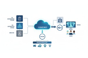
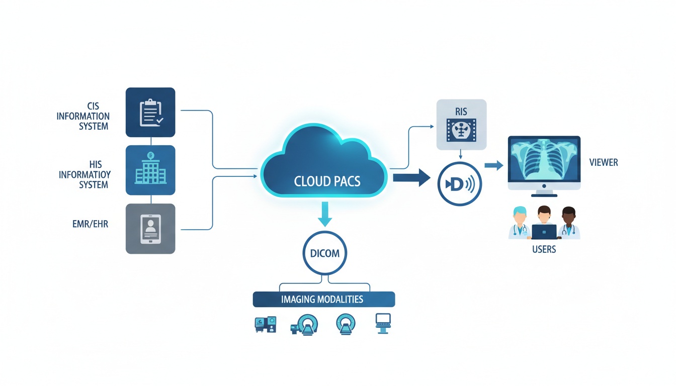
Regardless of the medical imaging method used, the images obtained can all be stored, retrieved, and distributed digitally within one integrated system – PACS (Picture Archive and Communication System), which comprises software to integrate data from different radiology imaging systems (such as CT, MRI, and X-ray), imaging information storage for archiving, retrieving images, and related documents, and a computer network connecting the system components and workstations for data access. The archiving, retrieval, and distribution within PACS are enabled by its transmission protocol – DICOM (Digital Imaging and Communications in Medicine).
The degree to which PACS has been able to simplify and speed up the management of digital patient imaging, improve workflow, and increase productivity has turned it into the functional backbone of modern radiology departments and diagnostic centers.
Another type of IT system commonly used in radiological practice is RIS (Radiology Information System), complementary to PACS and HIS (Hospital Information System), and usually used by radiologists for scheduling patients and tracking and interpreting examinations and billing, among other functions.
Clinical IT systems like PACS, RIS, and their protocols can be understood as the contemporary digital alternative for paper and film-based archiving in radiology, making the process more reliable and its management far less time-consuming. Recently this medical image archiving and processing technology has been made even more efficient and convenient by the development of cloud computing platforms like PostDICOM, eliminating the need for on-premises hardware within hospitals and other institutions employing radiology imaging.
PostDICOM takes the technology of conventional clinical IT systems a step further by enabling DICOM files to be stored in the cloud while preserving the functions of regular integrated systems relying on hardware.
This is something that we specialize in, and our PostDICOM service includes the following features:
 Storing any medical images (such as obtained from ultrasound diagnostic imaging, MRI, CT, and radiography) into DICOM format and storing them along with related clinical documents in PDF, JPG, BMP and AVI file formats in the cloud environment using HTML5 interface on browsers.
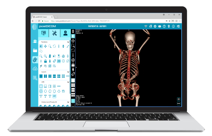
 Viewing files from desktop PCs, smartphones, and tablets through our sophisticated HTML5 zero footprint lossless DICOM viewer.
 Sharing files between doctors, medical groups, and patients.
 Storage – cloud space, with optional subscription fees for expansion of storage space.
 Security – secure storage and retrieval of patient data. Highest level of safety precautions is taken in order to prevent any system vulnerabilities.
 Advanced diagnostic tools such as MPR, MIP, MINIP, AVGIP, and 3D rendering and advanced image processing tools and data streaming algorithms.
PostDICOM software expands on the usage of clinical IT systems as archives and encourages exchange of ideas between users to facilitate consultations and interaction with patients.
|
Cloud PACS and Online DICOM ViewerUpload DICOM images and clinical documents to PostDICOM servers. Store, view, collaborate, and share your medical imaging files. |
 - Created by PostDICOM.jpg)