Have you ever wondered how doctors get such incredibly detailed insights into the inner workings of your body without a single incision? It’s not magic, but rather the marvel of modern medical imaging.
Today, we're going to pull back the curtain on one such powerful technique: SPECT imaging. If you've ever found yourself asking, "what is SPECT imaging?" or "how does a SPECT scan work?", you've come to the right place!
In the world of diagnostics, where precision is paramount, tools like SPECT (Single-Photon Emission Computed Tomography) scans play a crucial role. They help doctors detect diseases early, monitor treatment effectiveness, and understand the intricate functions of organs like the heart, brain, and bones.
Our goal today is to demystify this technology, making it easy to understand and grasp, without getting lost in complicated jargon.
At its core, SPECT imaging is a nuclear medicine procedure that uses a special type of camera and a tiny amount of radioactive material (called a radiotracer or radionuclide) to create 3D images.
Unlike X-rays or CT scans that show anatomical structures (like bones or organs), SPECT scans focus on function. They reveal how organs are working at a cellular level, showing blood flow, metabolic activity, and how tissues absorb or react to certain substances.
Imagine you want to know if a specific road in a city is congested. A regular map might show you the road, but it won’t tell you if cars are moving smoothly or stuck in traffic. SPECT is like a "traffic report" for your body, indicating activity levels and pinpointing areas of reduced or increased function.
This makes it invaluable for diagnosing conditions that affect how organs perform, often before structural changes are visible on other types of scans.
The process of a SPECT scan is fascinating and surprisingly straightforward from a patient's perspective. Let's break down "how does a SPECT scan work" step-by-step:
1. Introducing The Radiotracer: First, a small, safe amount of a radiotracer is injected into your bloodstream, or sometimes inhaled or swallowed, depending on the area being examined. This substance is specifically designed to travel to the organ or tissue of interest. For example, some tracers are absorbed by active heart muscle cells, while others target specific types of brain receptors or bone formations.
2. Tracer Distribution: Over a period of time (which can range from minutes to a few hours, depending on the tracer), the radiotracer travels through your body and accumulates in the target area. The amount of tracer that collects in an area is directly related to its activity or blood flow.
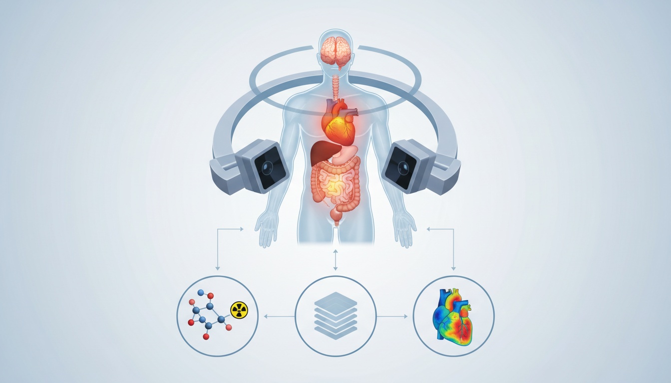
3. Emission Detection: Once the tracer has settled, you'll lie on a table that slides into the SPECT scanner. The scanner isn't an enclosed tunnel like an MRI; rather, it typically has one or more gamma cameras that rotate around your body. These cameras don't emit radiation; instead, they detect the tiny gamma rays emitted by the radiotracer within your body.
4. Image Reconstruction: As the cameras rotate, they capture images from many different angles. A powerful computer then takes all these 2D images and uses sophisticated algorithms to reconstruct them into detailed 3D cross-sectional slices of the organ or area being studied. This allows doctors to view the distribution of the radiotracer in three dimensions, providing a clear picture of how well an organ is functioning.
This entire process provides unique functional insights that other imaging modalities might miss, making SPECT a vital tool in modern medicine.
SPECT scans are incredibly versatile and are used across various medical specialties:
• Cardiology (heart): To assess blood flow to the heart muscle, detect coronary artery disease, evaluate damage after a heart attack, and determine the effectiveness of bypass surgery or angioplasty.
• Neurology (brain): To help diagnose and monitor conditions like Alzheimer's disease, Parkinson's disease, epilepsy, strokes, and even some types of brain injuries or tumors by looking at blood flow and metabolic activity.
• Oncology (cancer): While not a primary tool for initial cancer detection, SPECT can be used to locate certain types of tumors, determine if cancer has spread to bones, or evaluate the effectiveness of chemotherapy.
• Orthopedics/bone Scans: To detect bone infections, fractures (especially stress fractures that aren't visible on X-rays), and certain bone tumors.
• Infection Detection: In some cases, SPECT can help pinpoint the location of hidden infections.
This is a really common and excellent question: "spect vs pet scan" – what sets them apart? Both SPECT (Single-Photon Emission Computed Tomography) and PET (Positron Emission Tomography) are nuclear medicine imaging techniques that provide functional information about the body. They both involve injecting a radioactive tracer and detecting emissions from within the body to create images. However, the key difference lies in the type of radiotracer used and, consequently, the type of emissions they detect.
• Spect Scans: Use radiotracers that emit single gamma rays. The gamma cameras directly detect these gamma rays to create images. These tracers generally have longer half-lives (meaning they remain active for a longer period) and are often more readily available and less expensive.
• Pet Scans: Use radiotracers that emit positrons. When a positron encounters an electron in the body, they annihilate each other, producing two gamma rays that travel in opposite directions. The PET scanner detects these pairs of gamma rays simultaneously. PET tracers typically have shorter half-lives and often require an on-site cyclotron for production, making PET scanners more complex and generally more expensive to operate. The most common PET tracer is FDG (fluorodeoxyglucose), which helps visualize glucose metabolism, often highly active in cancer cells.
In summary:
| Feature | SPECT Scan | PET Scan |
| Tracer | Emits single gamma photons | Emits positrons (which then produce gamma rays) |
| Detection | Gamma cameras detect direct gamma emissions | Detects paired gamma rays from annihilation |
| Resolution | Generally lower resolution (but improving!) | Generally higher resolution and sensitivity |
| Cost/Access | Often more accessible and less costly to operate | Typically more expensive, often requiring special facilities |
| Information | Primarily blood flow, functional activity | Primarily metabolic activity (e.g., glucose use) |
Both techniques are powerful and often complementary. Sometimes, a doctor might even order both if different types of functional information are needed to get a complete picture.
 - Created by PostDICOM.jpg)
A very natural concern when undergoing any medical procedure is its safety. So, "is a SPECT scan safe?" The answer is generally yes, SPECT scans are considered very safe, but like all medical procedures, there are a few considerations.
The primary concern for many people is the exposure to radiation. It's important to understand a few points:
• Minimal Radiation Exposure: The amount of radioactive material (radiotracer) used in a SPECT scan is very small. The radiation dose from a SPECT scan is comparable to or often less than that from a conventional X-ray or CT scan, and the radiotracer quickly leaves your body through natural processes.
• Short Half-life: The radiotracers used have very short "half-lives," meaning they decay rapidly and lose their radioactivity quickly. This minimizes your radiation exposure.
• Allergic Reactions: Allergic reactions to the radiotracer are extremely rare. Most tracers are very well-tolerated.
• Pregnancy And Breastfeeding: If you are pregnant or suspect you might be, or if you are breastfeeding, it is crucial to inform your doctor. SPECT scans are generally avoided during pregnancy unless absolutely necessary, and special precautions might be advised for breastfeeding mothers to prevent the transfer of the tracer to the baby.
• Mild Side Effects (rare): While the actual "spect scan side effects" are minimal, some people might experience mild discomfort at the injection site (like a bruise or slight soreness). Rarely, very mild nausea or dizziness can occur, but these are usually fleeting.
The benefits of a SPECT scan in providing crucial diagnostic information far outweigh these minimal risks for the vast majority of patients. Your medical team will always weigh the benefits against any potential risks and discuss them with you.
In conclusion, SPECT imaging is a remarkable testament to how far medical technology has come. By providing a window into the functional world of our organs, it empowers doctors to make earlier, more accurate diagnoses and to tailor treatment plans with greater precision. It bridges the gap between seeing what is there and understanding how it's working, offering invaluable insights that can profoundly impact patient care.
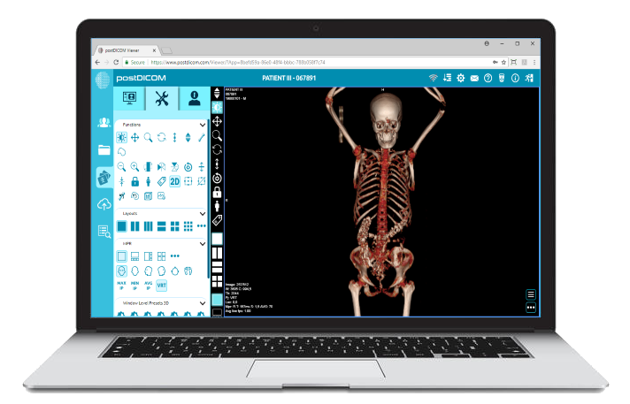
Understanding techniques like SPECT imaging helps you become a more informed participant in your own healthcare journey. And for healthcare providers, having access to clear, high-quality images and a robust system to manage them is absolutely essential.
Ready to enhance your diagnostic capabilities and streamline your workflow with cutting-edge medical imaging solutions?
Discover how PostDICOM's intuitive, powerful platform can transform your practice. Get started with a free trial today and experience seamless DICOM viewing, sharing, and archiving that truly makes a difference!
Click here to claim your Free Trial!
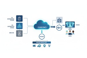
|
Cloud PACS and Online DICOM ViewerUpload DICOM images and clinical documents to PostDICOM servers. Store, view, collaborate, and share your medical imaging files. |