Routine imaging, emergency, or pain, swelling, trauma, or suspected cancer investigation are typically the scenarios that result in bone lesions being identified. Other lesions are harmless and remain stable over the years, whereas others can be an indication of an infection, metastatic, or primary bone tumors, which necessitate immediate treatment. Since management decisions rely much on the accuracy of imaging, radiology is central to evaluation.
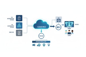
Here the DICOM standards play an imperative role. DICOM assists healthcare providers to store, share, compare, and analyze imaging studies in a consistent manner across hospitals, clinics, specialists, and imaging devices. That consistency can enhance the diagnosis, follow-up decisions, treatment planning, and collaboration in bone lesion radiology.
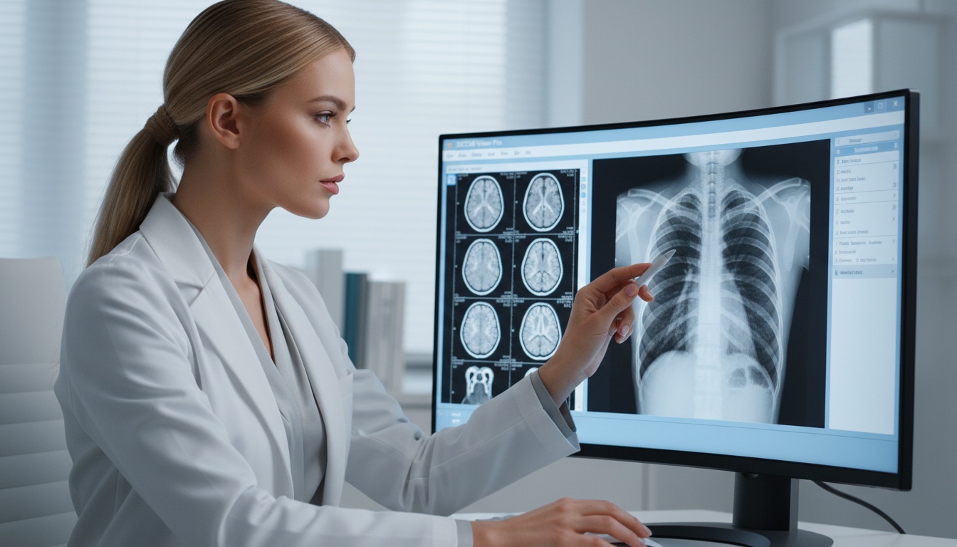
The DICOM standards facilitate the bone lesion radiology as they enable the storage and display of CT scans, MRI studies, X-rays, PET/CT images, and other imaging data in a standard format. This assists radiologists to compare previous examinations, concur with experts, track progression of lesions and make quicker and more precise clinical judgments.
Any defect in bone tissue visible on an imaging is considered a bone lesion. Bone lesions can be found either accidentally or when examining symptoms like pain, fracture, swelling or loss of movement.
Most bone lesions are benign such as bone cysts, fibrous dysplasia, enchondromas and healing abnormalities. There are other types which are aggressive or malignant including osteosarcoma, chondrosarcoma, multiple myeloma, or metastatic cancer.
Since appearances may be confusing, imaging interpretation is frequently the initial step in calculating urgency and further actions.
Subtle imaging results may be the difference between a benign lesion that is stable and an aggressive destructive lesion. Radiologists often evaluate:
• Lesion Margins And Zone Of Transition
• Cortical Thinning Or Destruction
• Periosteal Reaction
• Matrix Mineralization
• Soft Tissue Extension
• Pathologic Fracture Risk
• Growth Over Time
• Marrow Involvement
It is necessary to have accurate image access and comparison. In case previous research is not available or is not compatible, diagnosis might be deferred or unnecessary repeat testing requested.
DICOM is an acronym that means Digital Imaging and Communications in Medicine. It is the international standard when managing, storing, transmitting and displaying medical images.
DICOM is more than a picture-keeper. It also preserves critical metadata such as:
• Patient Identification
• Study Date And Time
• Modality Type
• Anatomical Region
• Slice Thickness
• Measurement Data
• Institution And Device Details
Such standardization enables images made by various manufacturers and healthcare facilities to collaborate more effectively.
Workups of bone lesions may include a series of imaging studies. A patient can start with X-rays and proceed to MRI, CT, PET/CT, biopsy planning, surgery, long-term surveillance.
Clinicians might find it difficult to compare studies or have full access to records without standardized workflows in imaging.
DICOM addresses most of these issues.
 - Presented by PostDICOM.jpg)
One of the most significant hints in assessing lesions is growth pattern. DICOM enables radiologists to access the previous imaging studies and see the difference with months or years.
An unchanged lesion can be considered in favor of a conservative monitoring. Quick development can mean the necessity to act immediately.
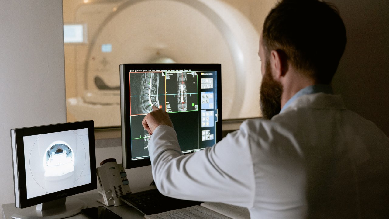
One modality is seldom used to evaluate bone lesions.
| Imaging Modality | Common Use in Bone Lesions |
| X-ray | Initial detection, matrix pattern, fracture risk |
| CT | Cortical bone detail, calcification, biopsy planning |
| MRI | Marrow involvement, soft tissue extension |
| PET/CT | Metabolic activity, staging |
| Bone Scan | Multifocal skeletal disease |
DICOM simplifies the process of reviewing these studies in a single workflow.
Complex bone lesions are usually referred to orthopedic oncology centers or tertiary hospitals. Compatible systems with DICOM enable transfer of imaging without loss of quality or failure of formats.
This will assist experts in examining cases more quickly and prevent scanning the same cases.
Multidisciplinary teams that might include many suspicious lesions are reviewed:
• Radiologists
• Orthopedic Surgeons
• Oncologists
• Pathologists
• Primary Physicians
DICOM image sharing facilitates collaborative decision-making and has a uniform image quality.
Accurate localization of lesions is of significance in terms of planning biopsy tracks, resections, fixation, or limb-sparing surgery. Access to standardized imaging enhances procedural confidence.
The management of bone lesions is not as effective when the imaging systems are scattered.
Cloud-based DICOM workflows help enable:
• Secure Image Sharing
• Faster Subspecialty Reads
• Clinician Remote Access.
• Multi-site Collaboration
• Enhanced Continuity Of Care.
This is particularly useful in rare or indeterminate bone lesions where expert opinion is important.
Modern healthcare increasingly relies on remote consultation. A musculoskeletal radiologist may review imaging from another city or country within hours.
Cloud-based DICOM workflows help enable:
• Secure Image Sharing
• Faster Subspecialty Reads
• Clinician Remote Access.
• Multi-site Collaboration
• Enhanced Continuity Of Care.
This is particularly useful in rare or indeterminate bone lesions where expert opinion is important.
Musculoskeletal imaging is a field in which artificial intelligence is become increasingly explored. Artificial intelligence tools might, in the future, help with:
• Lesion Detection
• Measurement Automation
• Follow-up Comparison
• Risk Prioritization
• Workflow Triage
Such systems rely on standardized, organized imaging data. DICOM is still the basis of a future AI-enabled radiology setting.
Modern cloud PACS platforms are beneficial to organizations that handle orthopedic, oncology, trauma, or outpatient imaging volume and facilitate workflows that are DICOM-native.
Benefits may include:
• Faster Study Access
• Centralized Archives
• Remote Collaboration
• Easier Referral Sharing
• Lower Infrastructure Burden
• Scalable Storage For Long-term Follow-up Imaging
In cases of bone lesions that could be under surveillance over years, good access to imaging is particularly important.
To enhance bone lesion imaging processes, organizations ought to take into account:
• Maintaining Dicom-compliant Archives
• Maintaining Past Research To Compare Over Time.
• Allowing Sharing Images Externally Securely.
• Reviewing With Multi-modality-friendly Viewers.
• Standardising Referral Imaging Workflow.
• Getting Infrastructure Ready To Integrate Ai.
The radiology of bone lesions is frequently a subject of close interpretation in time, modalities and specialities. It is seldom that one image tells it all. Comparing previous examinations, the collaboration with specialists, and the possibility to obtain all the imaging records can be very impactful.
That is possible with DICOM standards. DICOM assists medical institutions in efficiently, accurately, and teamwork management of cases of bone lesions by establishing a universal framework of medical imaging. With the ongoing transformation of the imaging ecosystem to cloud computing and artificial intelligence, the DICOM will be at the center.
X-rays are often the initial method, but MRI, CT, PET/CT or bone scans can be required based on the type of lesion, location, and clinical issue of interest.
No. Numerous bone lesions are harmless and might just have to be monitored. Imaging aids in deciding the necessity of additional examination.
Previously conducted studies assist radiologists to determine whether a lesion is stable, healing, or progressive.
Yes. DICOM standards are particularly created to provide interoperability in image exchange among compatible healthcare systems.
Cloud PACS enables remote access control, more convenient specialist consultation, and long-term control over images.
AI is being created to help with detection, measurement, and prioritization of workflow, but clinical use is not uniform across settings.
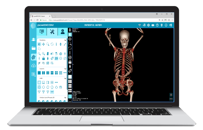
|
Cloud PACS and Online DICOM ViewerUpload DICOM images and clinical documents to PostDICOM servers. Store, view, collaborate, and share your medical imaging files. |