Over the years, Picture Archiving and Communication systems (PACS) have mainly been considered as medical image digital archives. Their task was simple, keeping radiology studies like CT scans, MRI scans and X-rays in their stores, and availing them to radiologists and clinicians. Although this is the main role, contemporary healthcare infrastructure has grown much more than mere storage.
Modern PACS systems are becoming more of a clinical data center, combining diagnostic imaging with other medical data and sophisticated analytics functions. The incorporation of electrocardiogram (EKG/ECG) analysis in the PACS systems is one of the most promising tasks in this evolution.
In cardiac monitoring, tremendous amounts of waveform data must be stored long term, structured analyzed, and quickly accessed by the clinician. Using the PACS infrastructure, healthcare providers will be able to centralize ECG data, automate the analysis of measurements, and provide sophisticated AI-based diagnostic assistance.
This change is part of a wider healthcare IT change: PACS is no more simply an image repository. It is emerging as a multi-modal diagnostic platform that can serve more complex clinical operations, such as advanced cardiology diagnostics.
Can PACS be used for advanced EKG analysis?
Yes. Current PACS systems are able to store, control, and examine ECG waveform data in standardized formats, e.g., DICOM waveform objects. PACS systems would facilitate advanced EKG analysis by:
• Concentrating On Ecg Data.
• Measuring And Detecting Abnormalities In The Heart, Automated.
• Combining Ecg Findings With Imaging And Patient History.
• Facilitating Long-distance Cardiology Visits.
• Facilitating Cardiac Diagnostics By Ai.
Consequently, PACS has the potential to serve as an all-encompassing cardiac diagnostic data system, instead of an imaging archive.
• Pacs Have Come Far Past Being An Image Storage Device With The Ability To Handle Multi-modal Clinical Data, Such As Ecg Waveform Data.
• Dicom Waveform Standards Allow The Storage Of Ecg Signals In Pacs, Which Can Be Integrated With Imaging Workflows.
• Pacs Platforms Can Utilize Automated Measurement Tools To Analyze The Ecg In Real-time And Detect Abnormalities.
• The Importance Of Pacs In Predictive Cardiology And Diagnostic Support Is Growing As A Result Of Ai Integration.
• Pacs Infrastructure Based In The Clouds Provides Cardiologists And Specialists With The Ability To Receive Ecg Data Remotely.
• Ecg Analysis Should Be Incorporated Into The Diagnostic Process To Enhance Diagnostic Efficiency, Collaboration, And Patient Care Outcomes.
PACS was initially meant to substitute film-based imaging archives. Physical storage of films used in hospitals in the past posed a challenge to the logistics of sharing and retrieval of studies. PACS revolutionized this process by making the imaging processes digital and allowing clinicians to electronically access studies.
Healthcare data grew more diverse, over time. Diagnostic data are now not only images, but also structured data, waveforms, lab data and patient monitoring data. The current state of healthcare thus demands systems that can process various forms of clinical data at the same time.
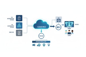
It is in this area that modern PACS systems have developed considerably. State-of-the-art PACS systems are now able to handle:
• Radiology Images
• Pathology Images
• Cardiology Imaging
• Ecg Waveform Data
• Clinical Reports
• Artificially Intelligent Diagnostic Data
Within this wider ecosystem, cloud-based PACS will be a central diagnostic ecosystem, facilitating the work of radiologists and cardiologists, as well as other specialists.
Electrocardiograms capture the electrical activity of the heart and are necessary in diagnosing a broad variety of heart disorders. Historically, ECG machines stored the data of the waveforms locally or sent it to special cardiology systems.
Nevertheless, such a strategy may disperse clinical data on several sites. By including ECG data in PACS infrastructure, providers will have a single location of imaging and cardiac diagnostic data.
Current PACS systems also allow the storage of ECG data as DICOM waveform objects, enabling the waveform signals to be saved in the same standardized format as medical images. This DICOM standard for medical imaging enables interoperability among ECG devices, cardiology systems, and imaging platforms.
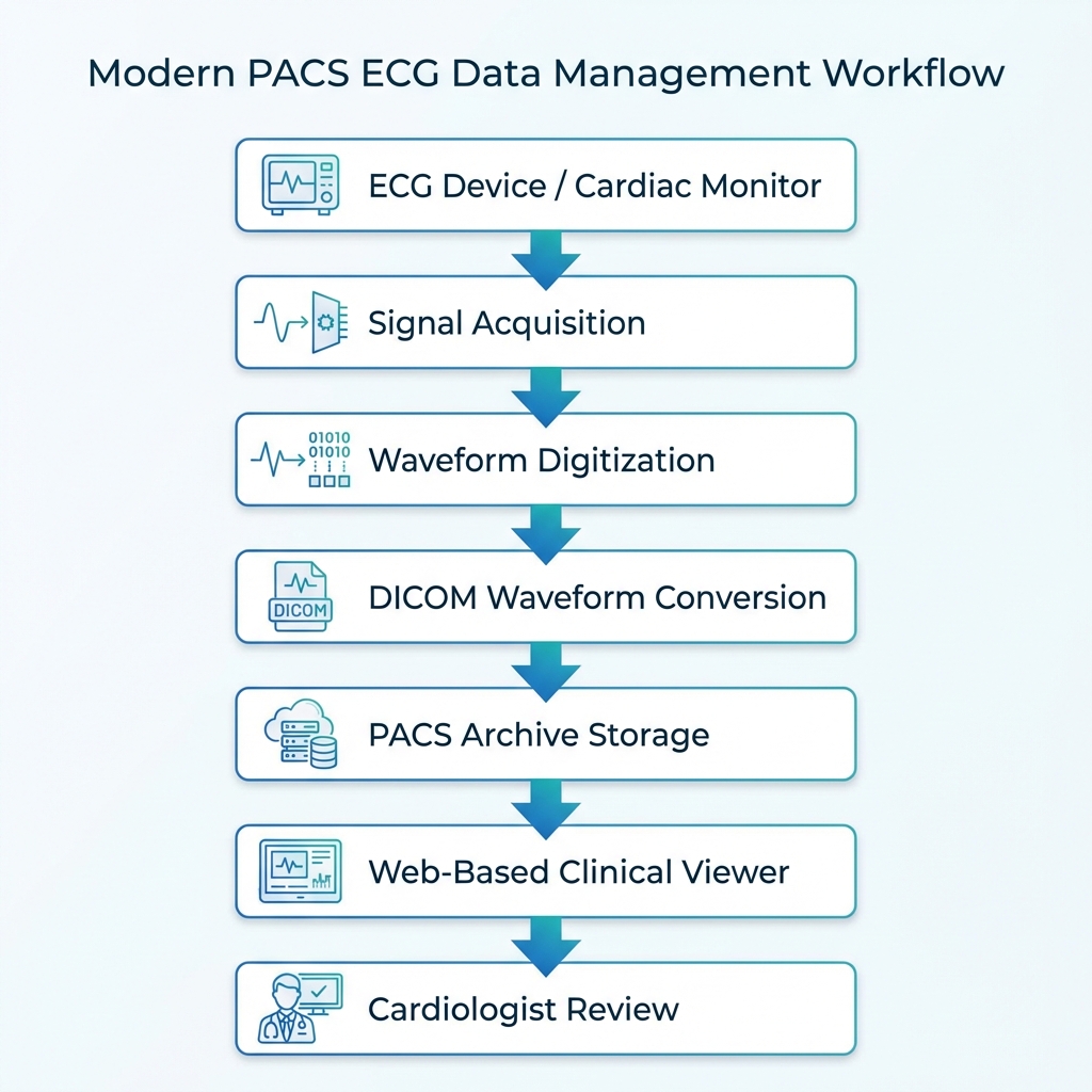
The standard process of ECG data within a PACS will involve:
1. Signal Acquisitionecg Devices Record Electrical Impulses In The Heart Of The Patient.
2. Digital Conversionthe Signals Are Computerized And Coded Into Systematic Waveform Data.
3. Dicom Waveform Encodingthe Data Of The Waveform Is Rearranged Into A Dicom Standard Format.
4. Pacs Storagethe Coded Waveform Is Stored On The Pacs System.
5. Clinical Accessthe Ecg Data Is Available To Cardiologists And Clinicians Via Pacs Viewers.
This integration helps ensure that cardiac data is stored alongside other diagnostic data, enhancing accessibility and continuity of care.
Automation of most areas of cardiac measurement and interpretation is one of the greatest benefits of incorporating ECG analysis in PACS.
Manual review of the ECG by clinicians has been a major part of traditional ECG interpretation. Although seasoned cardiologists are able to detect patterns within a short period of time, manual analysis remains time-consuming and erratic.
Contemporary PACS systems overcome this issue with automated measurement devices that can study ECG signals and estimate important cardiac measurements.
Examples of automated ECG measurements are:
• Heart Rate
• Pr Interval
• Qrs Duration
• Qt Interval
• St Segment Deviations
These automated systems are used to assist clinicians in quickly detecting abnormal patterns, which can signal cardiac diseases including arrhythmias, myocardial ischemia or conduction disorders.
With these measurements built into PACS workflows, clinicians get the ability to access ECG analysis results along with other diagnostic information to gain a better idea of the state of a patient.
 - Presented by PostDICOM.jpg)
Cardiology is one of the fields that are quickly changing with artificial intelligence. Machine learning algorithms are able to process ECG waveforms at a finer level of detail than a human observer can consistently do.
AI radiology algorithms can analyze ECG data to help detect small patterns that are related to cardiovascular diseases when implemented within PACS settings. These tools may help clinicians to identify possible abnormalities, prioritize the cases of highest need, and give insights on predicting cases.
AI-medical ECG analysis can assist in the identification of conditions such as:
• Atrial Fibrillation
• Ventricular Arrhythmias
• Myocardial Infarction Signs
• Heart Failure Risk Patterns
• Conduction Abnormalities
Implementing AI tools into the PACS infrastructure allows healthcare organizations to develop an integrated diagnostic environment where imaging data, waveform analysis, and machine learning insights can live side by side in the same clinical workflow.
This integration greatly improves diagnostic skills and minimizes the thinking load on clinicians.
Clinical workflow is also enhanced by the incorporation of ECG analysis in web-based PACS viewers. Clinicians can view imaging studies, cardiac waveforms and diagnostic reports on a single interface, rather than using several systems to review patient data.
This integrated access enhances the cooperation among health professionals. As an example, the ECG data can be analyzed together with the cardiac images ECG echocardiograms or CT angiography studies, which can be conducted by cardiologists. Diagnostic information can also be shared more effectively between radiologists and cardiologists.
An example of an integrated cardiology workflow could consist of:
 - Presented by PostDICOM.jpg)
When integrated into PACS infrastructure, these steps have helped healthcare organizations to optimize their diagnostic processes and minimize patient care delays.
Implementing ECG analysis into PACS settings has a number of significant benefits to healthcare and patients.
First, storing cardiac data centrally will make this information available consistently throughout the patients care journey. Clinicians have access to previous ECG data and imaging studies, allowing them to conduct longitudinal studies of cardiac health.
Second, automation increased efficiency. Computer-aided measurements and analytics can help to save time spent on the primary ECG interpretation and enable clinicians to concentrate on the more challenging diagnosis.
Third, integration of PACS enhances teamwork among clinical teams. The same diagnostic information can be provided to cardiologists, radiologists, and primary care physicians through a single system.
Lastly, PACS solutions based on the cloud computing system allow access to ECG data remotely. It improves multi-device DICOM viewer compatibility. Cardiac waveforms can be viewed by specialists practically anywhere, and it facilitates telecardiology services and better access to specialist care.
| Feature | Traditional ECG Systems | PACS-Integrated ECG Analysis |
| Data Storage | Local device storage | Centralized PACS archive |
| Accessibility | Limited access | Web-based access across departments |
| Workflow Integration | Separate cardiology systems | Integrated with imaging infrastructure |
| Automated Analysis | Limited | Advanced measurement tools |
| AI Integration | Rare | Increasingly common |
| Remote Access | Difficult | Supported through cloud PACS |
This analogy shows that PACS integration is changing the ECG analysis as a solitary diagnostic procedure to a joint clinic process.
However, on top of its benefits, incorporating ECG analysis into PACS infrastructure has a number of challenges as well.
Interoperability is one such consideration. Healthcare organizations will have to make sure that ECG equipment, PACS systems, and hospital information systems can communicate well. Standards like DICOM and HL7 are important in facilitating this integration, especially PACs integration with HIS and RIS.
The other issue is the data management. ECG waveforms produce huge amounts of time-series information which should be stored effectively yet without performance or accessibility degradation.
Security is a critical issue as well. Since ECG data includes sensitive information about patients, healthcare providers need to implement sound cybersecurity practices to secure medical records and ensure compliance with regulations.
Lastly, implementation has to be successful, which is impossible without clinical training. Medical workers should know how to apply the integrated PACS tools to obtain the full benefits of automated ECG analysis.
The role of the PACS in cardiology is set to grow as more and more healthcare systems are moving towards the use of cloud-based infrastructure. Cloud PACS solutions have scaling capabilities, remote accessibility, and high integration capacity to support advanced diagnostic processes.
Newer versions of PACS systems can introduce more advanced AI-powered models that forecast cardiac events before they manifest themselves. ECG data could be streamed into cloud PACS settings, and continuous monitoring devices and wearable sensors might transmit data on cardiac activity in real-time.
These advancements will make PACS a complete diagnostic ecosystem that incorporates imaging, waveform analysis, predictive analytics, and clinical decision support.
Cloud-based PACS for telemedicine are destined to take centre stage in the future of cardiovascular medicine by going beyond storage and adopting innovative data analysis possibilities.
Since its inception as a digital imaging repository, PACS technology has taken a new turn. Nowadays, the range of diagnostic functions facilitated by the medical imaging cloud platform is quite extensive, including advanced ECG data management and analysis.
Combining ECG waveform recording, automated measurements, and AI-based analytics, cloud PACS solutions can provide a more efficient cardiac diagnostics process and enhance collaboration between clinical teams. These features make PACS an integrated diagnostic architecture, but not a storage facility.
With the ongoing development of cloud-based healthcare solutions, the implementation of ECG analysis in the PACS setting is likely to grow more widespread. Cardiac diagnostics and patient care will be more effective and can happen faster and with greater accuracy when healthcare organizations adopt this evolution.
Yes. Current PACS software is capable of capturing the ECG waveform data in a DICOM waveform object, enabling cardiac signals to be stored along with medical imaging research.
By centralizing ECG data in PACS, clinicians may access cardiac waveforms more easily and enable automated analysis and review of cardiac waveforms together with imaging studies and patient records.
AI algorithms have the potential to process ECGs to identify any hidden anomaly, prioritize an emergency, and offer predictive results to assist a clinical decision-making process.
Yes. PACS systems based on clouds enable authorised clinicians to quickly access ECG information remotely via secure web-based viewers.
Yes. The inclusion of ECG analysis in the PACS infrastructure enhances the efficiency of the diagnostic process, interdisciplinary coordination, and simplifies the process of providing patients with comprehensive care.
|
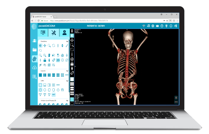
Cloud PACS and Online DICOM ViewerUpload DICOM images and clinical documents to PostDICOM servers. Store, view, collaborate, and share your medical imaging files. |