The internal organs and bones of our body are covered by skin and other tissue barriers, and are therefore not visible to the naked eye. The term ‘medical imaging’ is used to refer to techniques that allow us to view the interior of the body. This article will help you understand what is medical imaging and how it plays an important role in patient management today.
Diagnosis is the process of identifying a specific disease or illness based on a thorough examination of the patient. Unfortunately, most diseases and conditions affect areas of the body that are not normally visible to the naked eye. Diagnostic medical imaging can aid in diagnosis by allowing us to visualize any abnormalities that might exist within the body. For instance, in a patient who has sustained a trauma, medical imaging can tell us if any bones are broken or dislocated.
Diagnostic medical imaging relies on the use of ‘invisible’ waves, such as electromagnetic radiation, magnetic fields, or sound waves. Learning about these different types of waves help us to understand what is medical imaging science all about. The waves typically originate from a source placed on one side of the body, travel through the body (and through the region of interest), and hit a detector that is placed on the other side of the body. The waves are absorbed to varying degrees by different body tissues. This way the detector develops an image that is composed of ‘shadows’ of various body tissues. Earlier forms of medical imaging, such as radiographs, used a photodetector plate, which required film processing prior to visualization. Advanced medical imaging today allows images to be directly captured through a detecting camera and the images can be viewed digitally on a monitor.
Although a large part of medical imaging is performed mainly for diagnostic reasons, it has several other applications as well. A few of the most common applications of medical imaging are described below:
Spot diagnosis: As the name suggests, this is the most common application of diagnostic medical imaging. An image can tell us, at a glance, what exactly is wrong with the patient. Plain radiographs and CTs help detect fractures, cysts, tumors, and anomalies of the bone.
Monitoring disease progression: Diagnostic medical imaging is often used to determine disease stage and progression. In a patient with cancer, a contrast-enhanced CT or an MRI can be used to determine the exact stage of disease, while PET scans can detect any metastases. SPECT, a type of bone scan, has been found useful to monitor progression in Parkinson’s disease.
Treatment planning: Medical imaging also aids in treatment planning by allowing surgeons to determine the size of a lesion and hence the extent of surgery beforehand. Surgeons can perform virtual surgery using medical imaging technology, either directly in the software, or after importing and creating stereolithographic models.
Evaluating the efficacy of treatment: PET scans are often used in cancer patients undergoing treatment to check if the treatment regimen has been effective in diminishing the size of the tumor. Surgeons also use medical imaging during a surgical procedure to check if bones have been aligned properly or if implants have been placed in their proper position. Imaging can be done to assess long-term efficacy of treatment procedures. For instance, volumetric analysis of orbital contents is often performed six months after the procedure to check if orbital reduction and fixation after trauma was performed accurately.
Age-related calculations: Age can often be determined by assessing growth of internal body structures. For instance, fetal age and maternal gestational age are often determined through an ultrasound. Certain radiographs, such as hand-wrist and dental radiographs, are widely used to calculate a patient’s age if it is unknown or necessary for legal purposes.
|
Cloud PACS and Online DICOM ViewerUpload DICOM images and clinical documents to PostDICOM servers. Store, view, collaborate, and share your medical imaging files. |
There are several kinds of diagnostic medical imaging, depending on the physical nature of the waves employed and the method of image capture. There is no single imaging technology which is superior to the rest as each has its own advantages and disadvantages. Based on these limitations, radiologists today have found a specific ‘niche’ best-suited for each imaging modality:
As indicated by its name, ultrasound uses sound waves to acquire medical images. Since it does not involve electromagnetic radiation, it is probably the safest form of diagnostic medical imaging. The sound waves travel from the ultrasound probe through a conducting gel into the body. The waves then hit various anatomical structures inside the body and bounce back. They are captured and transformed into images that can be viewed on a monitor. A specialized form of ultrasound, called the Doppler, allows us to visualize the movement of blood within blood vessels.
Radiographs are the earliest form of medical diagnostic imaging. They are typically used to visualize bones and have largely been replaced by more advanced medical imaging systems. However, the traditional radiograph is still useful in a certain clinical situations:
Mammography: This is a radiograph of the breast. It is used as a screening tool in women to detect breast cancer.
Fluoroscopy: This technique uses radiographs in combination with a contrast agent that is either injected or swallowed. The path of the contrast agent is followed via radiographs to determine obstructions, ulcers, and other pathological processes.
In this technique, the patient lies within a CT chamber, which contains both the detector and the source. The source and detector lie opposite each other and travel in an arc around the patient, obtaining images serially. Images are taken in slices of a few millimeters each and in three different axes—producing coronal, axial, and sagittal sections. These sections can then be reconstructed to form a three-dimensional image. CT images possess far greater detail compared with traditional radiographs. However, CT scanning delivers a substantially higher dose of radiation to the body.
This diagnostic medical imaging technology makes use of radio-waves within a magnetic field. The human body is largely composed of water. When placed in the MRI scanner, the hydrogen ions within the water molecules align themselves according to the field. When radiofrequency waves are applied, this alignment changes and after that the ions return to their original position. These changes in alignment are recorded and processed to create an image. The MRI is useful for visualizing soft tissue structures such as muscles, tendons, and joint spaces. Although there is no radiation hazard, MRI can be dangerous for people who have metal implants because of the use of a strong magnetic field. This includes patients who have artificial joints, pacemakers, or other types of implants.
This technique involves the use of radioactive molecules that are called ‘tracers’. The tracers are either swallowed or injected into the bloodstream. Once within the body, tracers are taken up by specific tissues. The gamma rays emitted by these tracers are captured on a gamma camera and converted into digitized images. Tracers may be chosen based on the region of interest. For instance, imaging of the thyroid gland requires radioactive iodine, as this compound is preferentially taken up by thyroid cells. Bone scanning for infectious disease uses technetium, gallium or indium. Areas that take up the material will emit more radiation and will appear as ‘hot spots’ on acquired images.
A special type of nuclear imaging is positron emission tomography (PET). It can use a radioactive form of glucose. Glucose is preferentially taken up by cells which have a high rate of metabolism, such as cancer cells. Thus, this advanced diagnostic imaging technique can help to identify distant metastases in cancer patients.
As medical imaging continues to evolve, researchers are finding ways to improve diagnosis and treatment planning. One of the most exciting areas currently under research is the application of artificial intelligence (AI) to medical imaging. Artificial intelligence is the capacity of software or machines to replicate cognitive thinking exhibited by humans. They can therefore help in problem-solving tasks. AI in medical imaging can push new frontiers with regard to both diagnosis of diseases as well as planning and monitoring treatment efficacy. Following are some applications of AI in medical imaging:
Identifying slices of interest: A single CT or MRI scan of a patient can generate literally hundreds of images, as each slice is only a few millimeters in length. For the radiologist, going through each individual slice to detect abnormalities can be a very time consuming process. AI can be used to sift through all the slices and pick up only those slices that are of interest to the radiologist.
Detecting finer abnormalities: Very minor differences in color or contrast may not be visible to the naked eye. However, these differences may signal the early onset of invasive disease. AI can be used to pick up even minute differences, thus aiding in diagnostic accuracy that cannot be achieved by manual means.
Retrieving old records: AI can go through databases to retrieve older images from patients’ health records. These images can be used for comparison with any current images taken. This can be used for assessing disease progression or evaluation of the efficacy of treatment.
Large scale screening: A novel application of AI in medical imaging is large scale medical screening. A recent artificial intelligence based application was developed to screen medical images across multiple hospital databases. The AI was trained to detect large vessel obstruction, an early sign of stroke. If this works out, the application can alert the patient and the stroke specialist on a priority basis. It will reduce the time to treatment, which can significantly improve patient outcomes.
Preparing diagnostic reports: AI would be able to translate abnormalities in color and contrast into actual diagnostic findings. This could be done by feeding information based on earlier case records. Using diagnostic information, AI can also be used to generate imaging reports.
Medical images are after all just pictures. The better the quality of a picture, the more information it can provide. Keeping this in mind, the National Electrical Manufacturers Association (NEMA) released a standard, high quality format for viewing and storing medical images. DICOM, which stands for Digital Imaging and Communications in Medicine, is globally accepted. It cannot be accessed by ordinary computer programs. Special software applications, called DICOM viewers, are needed to view and edit modern-day medical images.
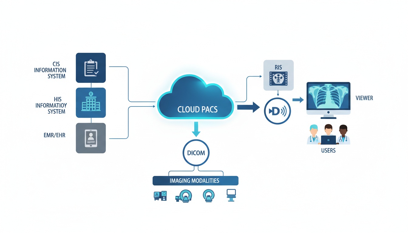
Since DICOM-based images are high quality and multiple images from a single patient scan require a lot of storage space, special arrangements must be made to store and retrieve images in the DICOM format. The database and server system that stores DICOM images is referred to as a PACS (Picture Archiving and Communication System). In general, each hospital has its own internal PACS server, and images acquired from patients in that hospital alone are stored there. The disadvantage of this is that patients who change hospitals for various reasons may not be able to access past images.
The introduction of cloud-based PACS has made viewing and accessing DICOM files a lot easier. Cloud technology allows DICOM files to be stored and processed via the internet. These files can be accessed from anywhere, using any device that has the required permissions and software. It simplifies accessing a patient’s medical records from different geographical locations.
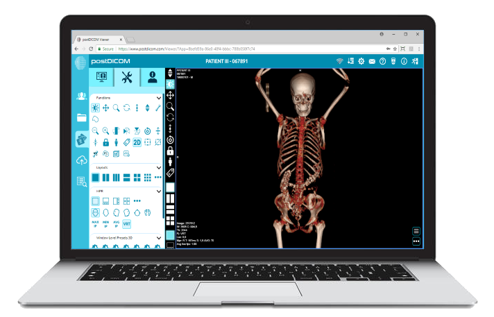
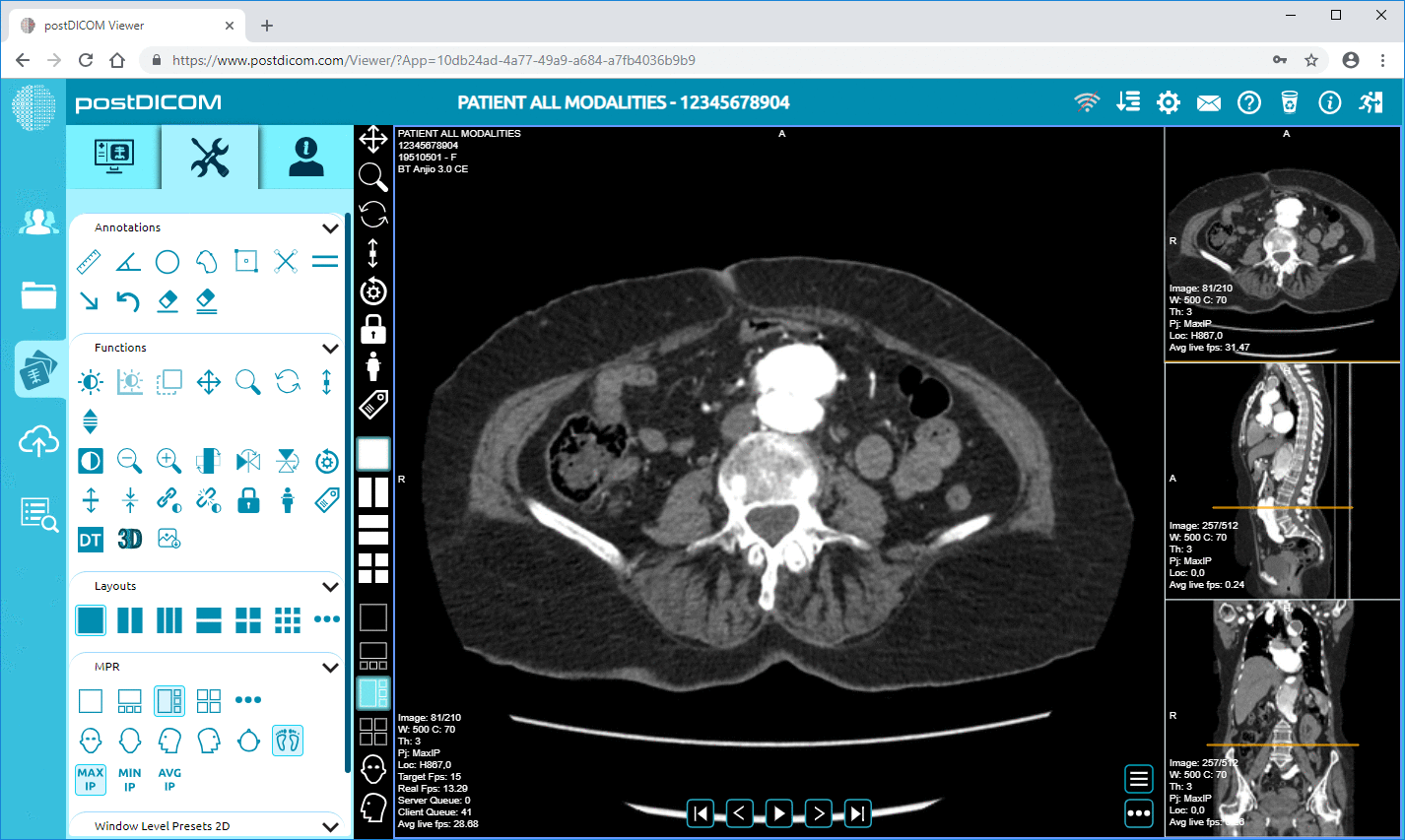
PostDICOM is an exciting, cutting-edge software application that meets the latest medical imaging technology demands. It is a smart DICOM viewer that not only helps you view medical images, it also offers advanced tools so that you can extract maximum information out of each image. These tools include three-dimensional and multiplanar reconstructed images, maximum and minimum intensity projections, and image fusion of two or more imaging modalities. PostDICOM is the only DICOM application that allows cloud-based image viewing. It is compatible with all operating systems, including Windows, iOS, Linux, and Android.
PostDICOM is for you to use — so give it a try today! You can expand the cloud storage space for a nominal fee.
|
|
Cloud PACS and Online DICOM ViewerUpload DICOM images and clinical documents to PostDICOM servers. Store, view, collaborate, and share your medical imaging files. |
 - Created by PostDICOM.jpg)