When it comes to diagnosing and evaluating heart disease, medical imaging is no longer a luxury—it's a necessity. Cardiac conditions often present with subtle symptoms, or worse, no symptoms at all until it's too late. That makes early and accurate imaging one of the most powerful tools in the arsenal of cardiologists, radiologists, and clinicians alike. Two of the most advanced imaging technologies for heart evaluation are Cardiac MRI (Magnetic Resonance Imaging) and Cardiac CT (Computed Tomography). Both are widely used. Both are highly informative. But they are not the same.
So the question often arises: Which one is better? The answer isn’t as simple as picking a winner. Cardiac MRI and CT each have strengths that make them uniquely valuable depending on what you're trying to see, measure, or confirm.
In this blog, we'll explore what each scan does best, when and why a doctor might prefer one over the other, and what kind of insights each one offers that the other can’t. We’ll also explore how imaging data from these scans is handled and reviewed in clinical workflows, with platforms like PostDICOM offering cloud-based tools that support faster, collaborative, and more accurate interpretation.
A Cardiac MRI is a non-invasive scan that uses strong magnetic fields and radio waves to produce detailed, high-resolution images of the heart. Unlike X-rays or CT scans, it does not use ionizing radiation, which makes it a safer option for patients who require repeated imaging or long-term monitoring.
Cardiac MRI is especially useful for evaluating soft tissue structures and functional aspects of the heart. It provides clear, layered images of the myocardium (heart muscle), valves, pericardium, and blood chambers. It's also capable of assessing cardiac motion, ejection fraction, and wall thickness with incredible precision. In fact, it is often considered the gold standard for measuring cardiac volumes and function.
Cardiac MRI excels in identifying conditions such as:
• Myocarditis And Other Forms Of Inflammation
• Cardiomyopathies (dilated, Hypertrophic, Restrictive)
• Pericardial Disease
• Myocardial Infarction And Scarring
• Congenital Heart Disease
It also allows for tissue characterization, which means clinicians can see signs of edema, fibrosis, or infarcted tissue using contrast-enhanced sequences like Late Gadolinium Enhancement (LGE). These are things a CT scan cannot easily detect.
A Cardiac CT, or more specifically a Coronary CT Angiography (CCTA), is a rapid, highly effective imaging test that uses X-rays to create detailed images of the heart's structures, particularly the coronary arteries. It is often used in cases of suspected coronary artery disease (CAD) to detect blockages or narrowing in the vessels supplying blood to the heart.
The real power of cardiac CT lies in its speed and clarity when it comes to visualizing the coronary arteries. With the help of contrast agents, CT scans can clearly map the arterial lumen and walls, identify atherosclerotic plaques, and even detect calcifications, which are early indicators of coronary disease.
Cardiac CT is typically used for:
• Coronary Artery Calcium Scoring
• Ruling Out Cad In Low-to-intermediate Risk Patients
• Evaluating Chest Pain In Emergency Settings
• Pre-operative Planning For Valve Replacements Or Bypass Surgery
Because CT imaging is fast—taking just a few seconds—it’s ideal for emergency diagnostics and high-throughput settings. However, it does expose patients to ionizing radiation and often requires the use of iodinated contrast, which can be problematic for those with kidney dysfunction or contrast allergies.
This is where the comparison gets technical. When evaluating coronary arteries, CT scans are clearly superior. They offer better spatial resolution, can visualize calcified and non-calcified plaques, and allow clinicians to detect even minor stenosis (narrowing) that could lead to heart attacks.
MRI, on the other hand, is more suited for evaluating larger vessels and vascular structures outside the coronary arteries. It's used in congenital conditions that affect the aorta, pulmonary arteries, or systemic venous return. In such cases, MRI offers a broader field of view and deeper tissue characterization without radiation.
So while CT is better for coronary arteries, MRI is better for vessels where you need more functional or tissue-level information. Each has a distinct role, and choosing the right modality depends heavily on the clinical question being asked.
Let’s put them side by side to see how they compare in some of the most important categories clinicians care about:
• Speed: CT wins. A full cardiac CT takes seconds. MRI scans often take 30-60 minutes.
• Radiation: MRI has none. CT uses ionizing radiation.
• Soft Tissue Detail: MRI is far superior, offering detailed views of heart muscle and tissue composition.
• Coronary Artery Imaging: CT is best for this, especially for detecting calcified plaque and stenosis.
• Contrast Safety: MRI uses gadolinium, which is less nephrotoxic than the iodine used in CT, but gadolinium isn’t risk-free either.
• Patient Comfort: CT is quicker and less likely to cause claustrophobia. MRI requires patients to lie still longer in a narrower tube.
• Implant Compatibility: CT can scan patients with most implants; MRI requires specific conditions to be met for safety.
The bottom line? Use CT when speed and coronary detail are critical. Use MRI when functional assessment and soft tissue imaging are the priority.
Cardiac MRI offers insight into the tissue health and function of the heart in ways that CT simply cannot. For instance, after a heart attack, it can assess the viability of heart muscle—a crucial piece of information in deciding whether to proceed with revascularization. CT might show a blockage, but only MRI will tell you if the downstream tissue is dead or salvageable.
MRI can detect:
• Fibrosis(scar Tissue)
• Edema(swelling Due To Inflammation Or Acute Injury)
• Perfusion Defectsduring Stress Imaging
• Myocardial Strain And Motionabnormalities
• Pericardial Thickening Or Effusion
It also doesn’t stop at anatomy. MRI lets you measure blood flow dynamics and perform tissue tagging, which gives you a clear view of how well each segment of the heart is contracting.
For structural heart disease, inflammatory conditions, and cardiomyopathies, MRI is often the imaging modality of choice.
 - Created by PostDICOM.jpg)
The choice between MRI and CT is almost always driven by clinical context. Here are some common considerations:
• Suspected Coronary Artery Disease: CT is faster, cheaper, and highly effective for ruling out blockages. It’s the default choice unless contraindications exist.
• Structural Or Functional Heart Disease: MRI provides more comprehensive data on heart muscle, valves, and tissue abnormalities.
• Emergency Settings: CT wins here due to speed and wider availability.
• Radiation Concerns: MRI is preferable for young patients, pregnant women (after first trimester), and those requiring repeated scans.
• Renal Impairment Or Contrast Allergies: Gadolinium-based contrast used in MRI is generally safer for kidneys but still used cautiously.
• Implants And Devices: CT is used if MRI is contraindicated due to pacemakers or defibrillators (unless they're MRI-safe).
In practice, many patients undergo both scans over the course of evaluation and treatment. Each modality contributes pieces to the diagnostic puzzle. The decision ultimately comes down to the diagnostic goal, patient safety, and imaging availability.
Cardiac MRI and CT are not competitors—they are complementary. Each offers a unique lens through which clinicians can view the heart, its structure, function, and vascular health. CT is the go-to for rapid, precise imaging of coronary arteries, perfect for acute evaluations and rule-outs. MRI is the standard for deep tissue insight, motion analysis, and understanding myocardial viability.
For patients and clinicians alike, the goal is not to choose the "best" scan, but the right scan at the right time.
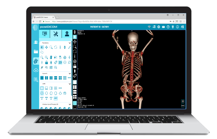
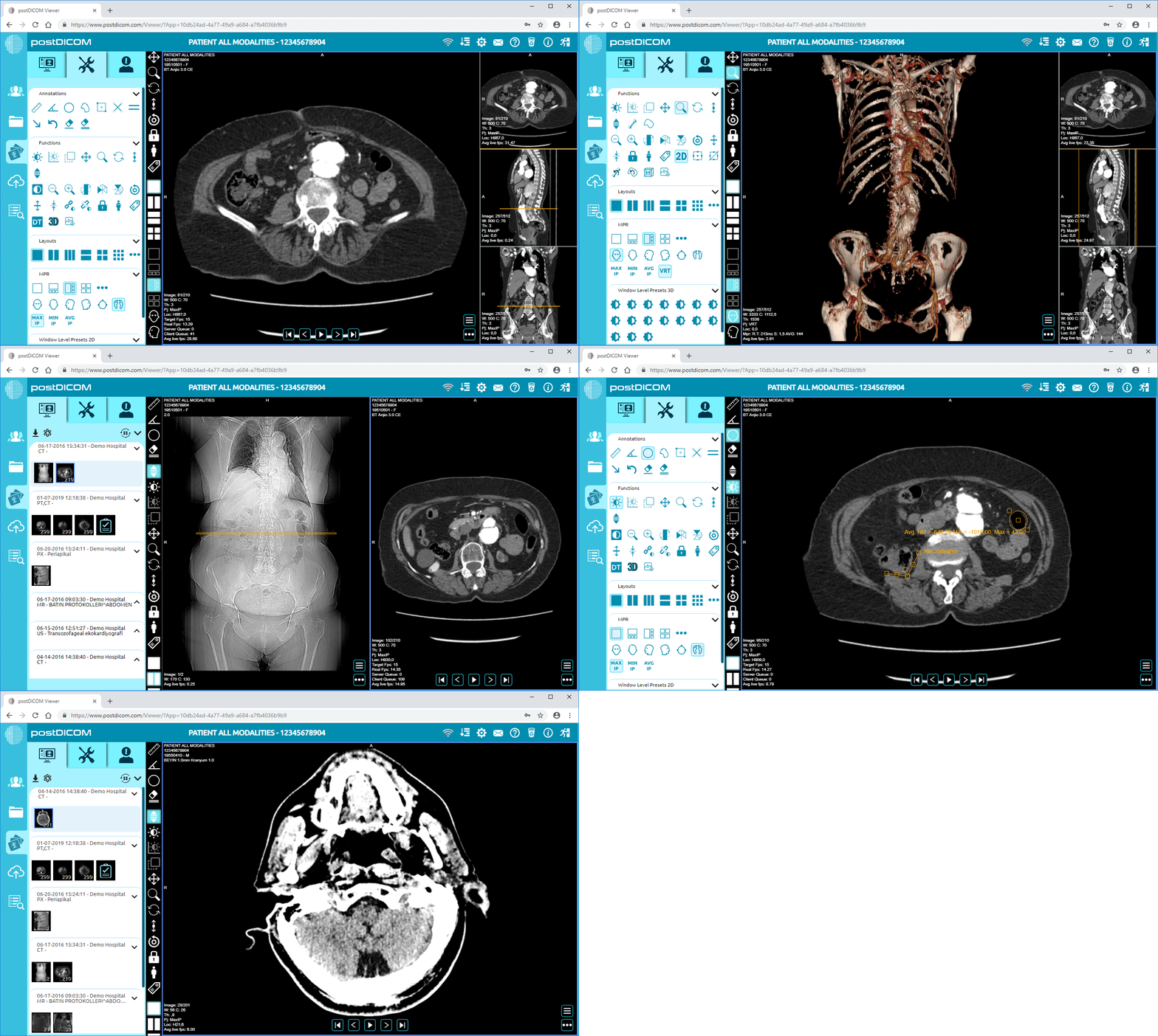
In a clinical environment where imaging data comes from multiple sources and systems, having the right tools to manage and interpret these scans is critical. That’s where PostDICOM comes in. With PostDICOM’s cloud-based viewer, healthcare teams can upload, review, and collaborate on both cardiac CT and MRI scans, complete with annotations, DICOM tag access, and secure sharing.
Whether you're preparing for surgery, investigating chest pain, or monitoring treatment response, your heart imaging workflow deserves more than just storage—it deserves intelligence, speed, and simplicity.
Start your free trial of PostDICOM today and bring your cardiac imaging into the future.


|
Cloud PACS and Online DICOM ViewerUpload DICOM images and clinical documents to PostDICOM servers. Store, view, collaborate, and share your medical imaging files. |