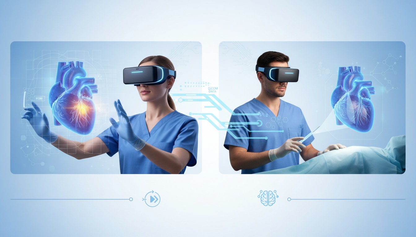
Picture this: A surgeon, donning a sleek headset, steps into a virtual realm where the intricate pathways of a patient's heart come alive in 3D. With a simple gesture, she rotates the heart, zooms into a problematic valve, and plans her surgical approach with unprecedented precision.
This isn't a scene from a sci-fi movie but a glimpse into the present-day medical world, where the boundaries of DICOM visualization are being pushed like never before.
Medical professionals have relied on flat, two-dimensional images for years to make sense of complex anatomical structures. But as the saying goes, "A picture is worth a thousand words, but experience? That's priceless."
With the advent of technologies like virtual reality (VR) and augmented reality (AR), medical imaging is undergoing a seismic shift, offering experiences that are immersive, interactive, and incredibly insightful.
As we explore advanced visualization techniques, we'll dive deep into the transformative potential of VR and AR in DICOM data interpretation.
From enhancing surgical precision to revolutionizing medical training, these technologies are changing how we see, understand, decide, and act in healthcare.
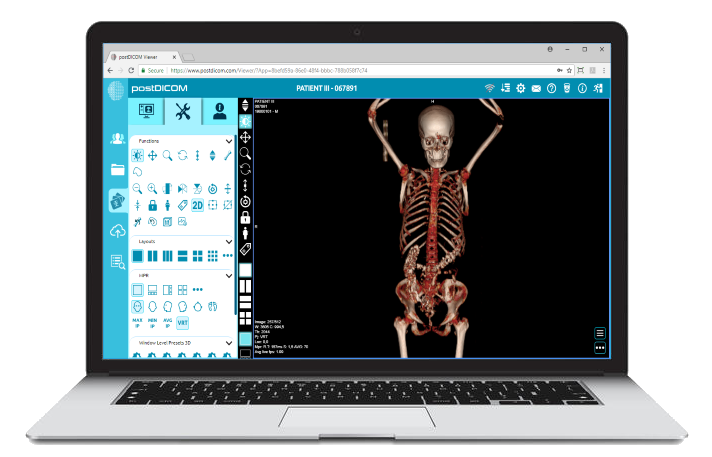
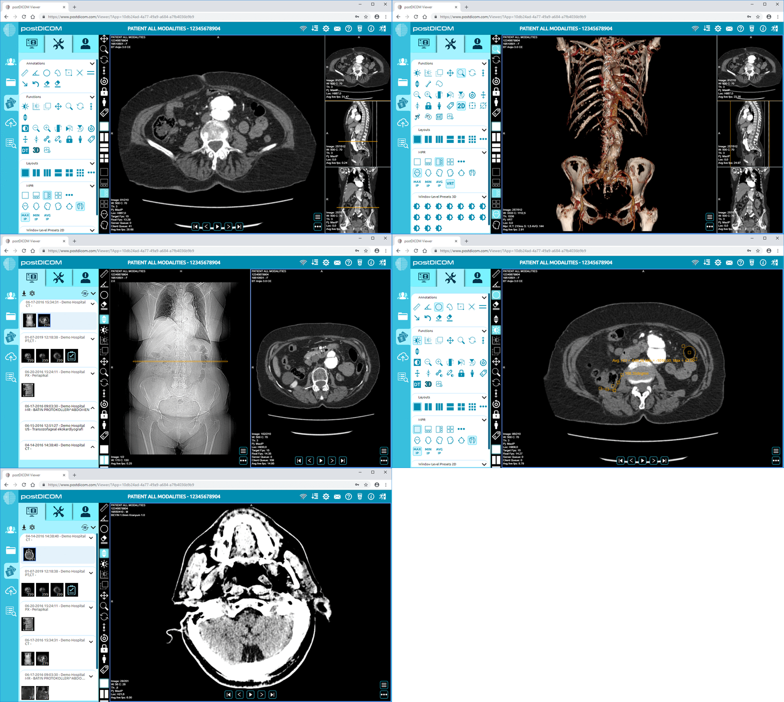
DICOM data visualization has been confined to two-dimensional images on computer screens for decades. Radiologists and medical professionals would sift through stacks of images, often cross-referencing multiple views to comprehensively understand a patient's anatomy.
While these 2D images have been instrumental in countless diagnoses and treatments, they offer a limited perspective, especially when understanding the spatial relationships between anatomical structures.
The human body, with its intricate web of tissues, organs, and vessels, is a marvel of complexity. When visualized in 2D, specific structures can overlap, obscure, or appear deceptively similar to adjacent tissues. This can pose significant challenges, especially in cases where precision is paramount.
For instance, planning a surgical procedure or pinpointing the exact location of a tumor requires a depth of understanding that 2D images might not always provide. While minimized with expertise and experience, the risk of misinterpretation still lingers.
More detailed and immersive visualization techniques are needed as medical procedures and treatments have evolved. Consider the case of a neurosurgeon navigating the dense network of the brain or an orthopedic surgeon planning a joint replacement.
A flat image fails to convey the required depth and detail in such scenarios. The need for a more 'tangible' and 'navigable' representation of DICOM data has become increasingly evident, paving the way for innovations in visualization.
Virtual Reality, often associated with gaming and entertainment, has made a groundbreaking entry into the medical world. By donning a VR headset, medical professionals can enter a virtual space where DICOM data comes alive in three dimensions.
It's as if they are walking inside the human body, witnessing its wonders up close. This immersive experience offers a depth of understanding that traditional methods simply cannot match.
With VR, DICOM data is no longer confined to flat screens. Complex structures can be viewed from all angles, rotated, zoomed in, or virtually dissected. Imagine a cardiologist being able to traverse the chambers of a heart or an oncologist pinpointing the exact boundaries of a tumor.
Such detailed visualization aids in accurate diagnosis, meticulous treatment planning, and even patient education, where individuals can 'see' their medical conditions in an entirely new light.
The implications of VR in DICOM visualization extend beyond diagnostics. Medical training, for instance, is witnessing a revolution. Medical students can explore virtual anatomical models, gaining hands-on experience without the constraints of real-world scenarios.
For surgeons, VR offers a rehearsal platform. They can simulate surgeries, practicing their approach and refining their techniques before the actual procedure, thereby reducing risks and improving outcomes.
The theoretical benefits of VR in DICOM visualization are being realized in clinics and hospitals worldwide. For instance, a neurosurgery unit in Europe uses VR to map out intricate brain surgeries, ensuring minimal damage to healthy tissues.
In another case, a rehabilitation center in Asia employs VR to help stroke patients visualize and understand their brain injuries, aiding their recovery process. These real-world applications underscore the transformative potential of VR in enhancing patient care and medical outcomes.
While Virtual Reality immerses users in a completely digital environment, Augmented Reality (AR) seamlessly blends the digital with the real world.
Through AR glasses or devices, medical professionals can overlay DICOM data onto the physical environment, creating a fusion of imagery that offers a unique perspective.
Imagine a surgeon viewing a patient's internal anatomy in real-time during a procedure, with DICOM data superimposed to guide each move. That's the magic of AR.
One of the standout benefits of AR in DICOM visualization is its potential for real-time decision-making. During surgeries or interventions, doctors can access and view DICOM data without diverting their attention from the patient.
This on-the-fly access to crucial information can be invaluable, especially in complex or emergency scenarios where every second counts. The ability to juxtapose digital imagery with the real world ensures that medical decisions are informed, precise, and timely.
Beyond the operating room, AR plays a pivotal role in patient engagement and education. Using AR devices, patients can 'see' their medical conditions, understand their anatomy, and grasp the implications of potential treatments.
This visual and interactive approach demystifies medical jargon, empowering patients to participate actively in their healthcare journey.
Additionally, AR facilitates collaborative diagnostics. Medical teams can collectively view and discuss DICOM data in a shared augmented space, fostering collaborative decision-making and holistic patient care.
The theoretical promise of AR is being actualized in medical facilities across the globe. In a renowned orthopedic clinic in North America, surgeons use AR to guide joint replacement surgeries, ensuring optimal alignment and fit.
Meanwhile, a pediatric unit in Australia employs AR to explain complex cardiac conditions to young patients and their families, making the information accessible and less intimidating.
These instances highlight how AR enhances medical procedures and transforms the patient experience when combined with DICOM data.
While integrating VR and AR with DICOM data offers immense potential, it's not without its challenges. The sheer volume and complexity of DICOM data demand robust computational power for smooth VR and AR experiences.
Latency, resolution limitations, or software incompatibilities can hinder the seamless visualization that medical professionals rely on. Ensuring these advanced visualization tools are accurate and responsive is paramount, especially in critical medical scenarios.
Beyond the technical aspects, there are ethical and practical considerations to navigate. How do we ensure patient data privacy in shared AR environments? How do we balance providing immersive experiences without causing sensory overload or discomfort to users?
Training medical professionals to use these tools effectively while ensuring they don't become overly reliant on them at the expense of their expertise is a delicate balance.
Despite these challenges, the road ahead is promising. Continuous advancements in technology are addressing many of the current limitations.
For instance, developing lightweight AR glasses with better battery life and higher resolution can enhance the user experience.
On the software front, AI-driven algorithms are being integrated to offer real-time insights and analytics during DICOM data visualization, making the process more intuitive and insightful.
As we look to the future, the convergence of VR, AR, and DICOM data is set to redefine the boundaries of medical imaging. We might see fully interactive holographic DICOM data displays, remote AR-guided surgeries where experts from across the globe collaborate in real time, or even patient-specific VR simulations to predict medical outcomes.
The fusion of technology and medicine is paving the way for a future where diagnostics, treatments, and patient care are more precise, immersive, and patient-centric.
The realms of DICOM visualization, once confined to flat screens and traditional methods, are expanding into exciting territories with the advent of VR and AR. As we've journeyed through the transformative potential of these technologies, it's clear that the future of medical imaging is not just about seeing but experiencing.
While challenges persist, the synergy of technology and medical expertise promises a horizon where diagnostics are more immersive, treatments more precise, and patient care more holistic.
As we stand at this intersection of innovation and healthcare, one thing is certain: the future of DICOM visualization is not just bright; it's revolutionary.


|
Cloud PACS and Online DICOM ViewerUpload DICOM images and clinical documents to PostDICOM servers. Store, view, collaborate, and share your medical imaging files. |