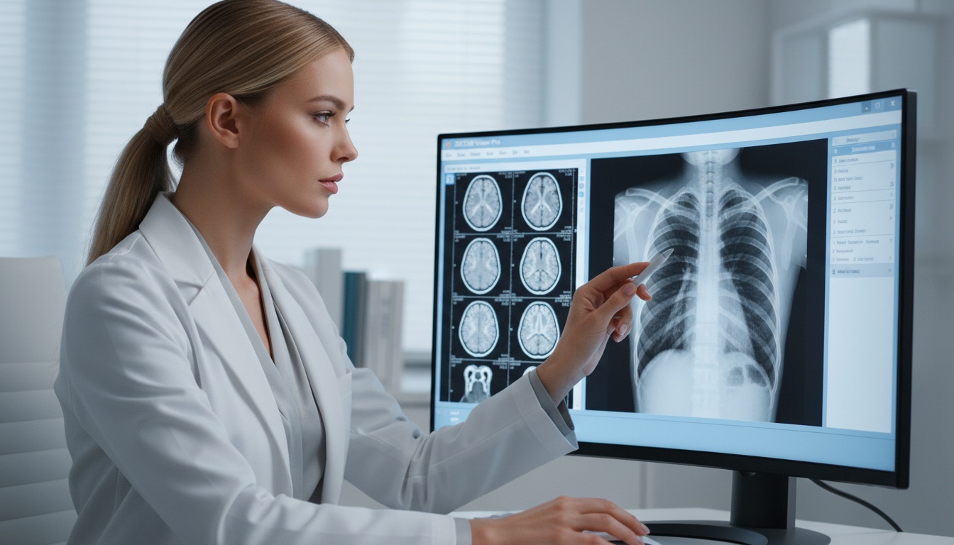
The era of X-rays on film and black-and-white images is a distant memory. Healthcare professionals now demand quicker access, better clarity, and tools to better comprehend complex anatomy. As medicine evolves, the future of digital imaging is not just better quality scans - it's a better way to view them.
Picture a surgeon exploring a CT scan in a fully immersive 3D environment prior to surgery. Imagine a radiologist working with remote experts to interactively manipulate models of the anatomy. Such scenarios are becoming a reality with new visualization tools rendering DICOM imaging data.
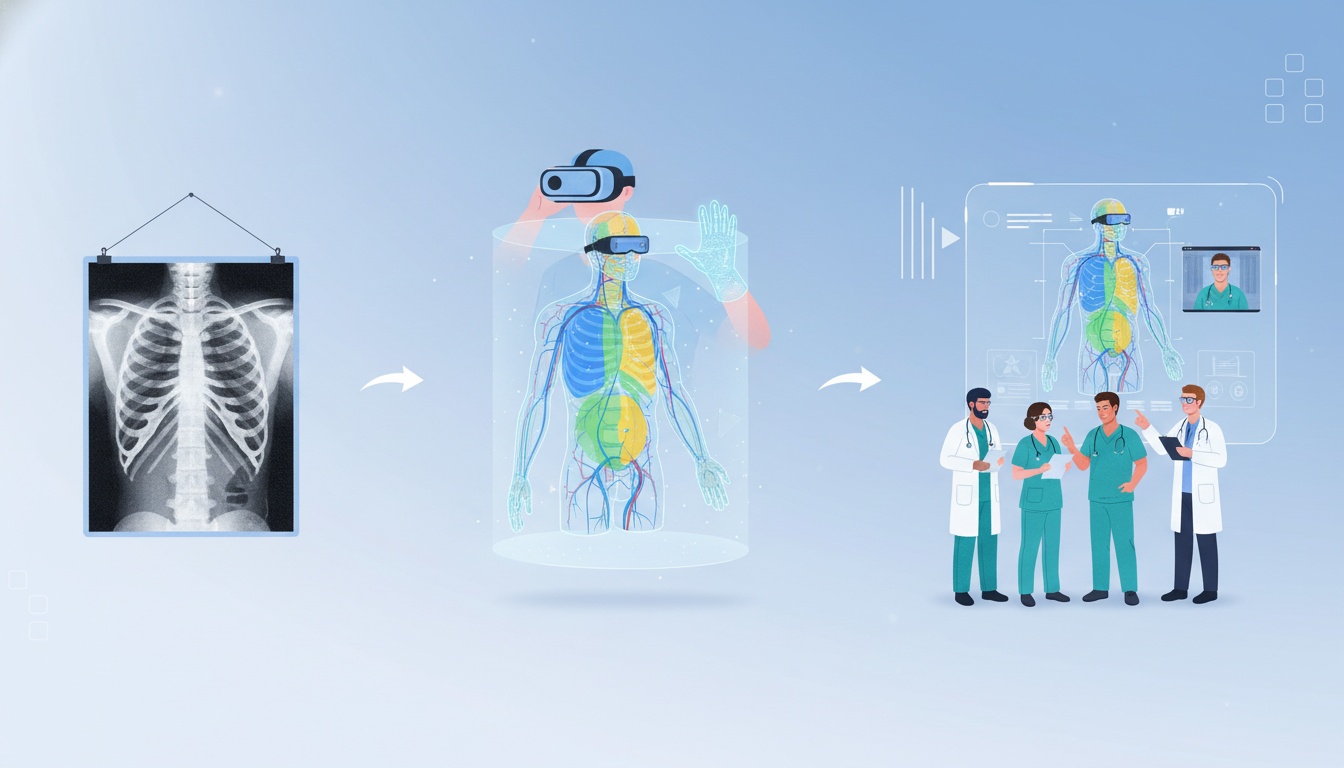
Virtual reality (VR), augmented reality (AR) and superior 3D rendering are on the horizon for radiology, surgery, medical training and telehealth. As this technology advances, health care organizations that upgrade their imaging workflows now will be well-prepared for the immersive world of the future.
DICOM advanced visualization tools convert routine CT, MRI, ultrasound and other medical studies into interactive 3D volumes that are more intuitive to comprehend and communicate. These techniques include volume rendering, multiplanar reconstruction (MPR), maximum intensity projection (MIP), virtual reality (VR) and augmented reality (AR). Rather than viewing only 2D slices of images, doctors can view anatomy in 3D, streamline planning for surgery, foster collaboration and better communicate complex imaging results.
DICOM (Digital Imaging and Communications in Medicine) is the international standard for image archiving, communication and presentation. It's used to communicate between imaging equipment like CT, MRI, ultrasound and PACS systems.
For decades, the standard way that most clinicians viewed images was 2D - one slice at a time on a desktop computer. Although this approach remains critical, many cases now involve large datasets with hundreds or thousands of images. It can be difficult to review these cases efficiently when there are complex spatial relationships.
This has led to a shift toward cutting-edge visualization tools to improve the interactivity, intuitiveness and utility of imaging.
Traditional 2D viewing is the bread-and-butter of radiology, but other specialties require more. Surgical, oncological, cardiovascular, orthopedic and emergency medicine specialists frequently want to understand the relationships between structures in 3D space.
That is where advanced visualization adds value. Rather than imagine how structures come together, they can explore 3D models. This may help avoid interpretive bottlenecks, speed up treatment planning conversations and build confidence in complex decisions.
With an ever-increasing volume of imaging studies, medical providers are looking for ways to be more efficient without compromising quality.
Multiplanar Reconstruction (MPR) enables reformatting data into coronal, sagittal, oblique and other planes. It enables clinicians to view anatomy from different angles without having to re-scan the patient.
MPR is widely used in spine imaging, orthopedic review, abdominal imaging, and vascular studies.
MIP creates an image from the brightest voxels in a volume, and is particularly well-suited to angiography and vascular studies.
It can be used to better image blood vessels, calcifications and contrast-enhanced structures.
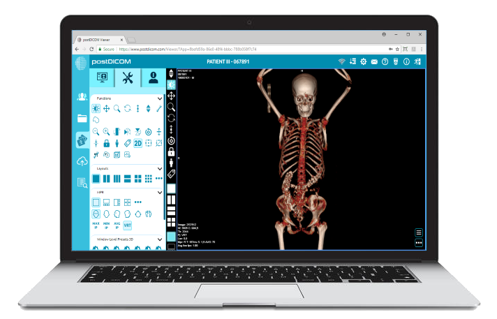
Volume rendering transforms images into interactive 3D models that can be rotated, zoomed, segmented and explored in real time.
It's useful for surgical planning, trauma evaluation and sophisticated anatomical analysis.
Modern systems can segment the lungs, tumors, fractures, blood vessels, or implants. Quantification can aid measurements, therapy planning, and follow-up.
Virtual reality is an immersive digital environment in which clinicians can view and interact with the patient anatomy via headsets and motion controllers.
Instead of looking at a 2D image on a computer, they can "walk" through the data and explore anatomy. This could help with understanding complex anatomy in surgical or multidisciplinary case discussions.
Potential benefits include:
• Better Depth Perception
• More Intuitive Anatomy Review
• Improved Surgical Rehearsal
• Improved Education And Simulation
• Greater Engagement During Case Conferences
With hardware costs dropping and software becoming easier to use, VR is set to become even more important in imaging.
Augmented reality (AR) involves superimposing virtual imaging data on the real world.
A surgeon may see anatomical information during surgery or a teacher may project interactive anatomy models in a classroom or lab.
Potential AR applications include:
• Procedure Navigation Support
• Image-guided Interventions
• Training And Education
• Improved Patient Communication
• Real-time Anatomy Reference During Planning
AR can bridge the gap between imaging data and real-world clinical action.
Virtual and augmented reality are increasingly used in medical care, but take-up depends on speciality and financial and workflow readiness. Academic and surgical programs, innovation-driven hospitals and advanced imaging departments tend to be the first to use it.
The near-term opportunity for many is not to replace existing radiology workstations. Rather, the opportunity is to use immersive visualization sparingly for complex surgical cases, multidisciplinary case planning, education and patient communication. Improvements in the efficiency and cost of hardware and software ecosystems will lead to increased adoption in the coming years.
VR and AR are moving from the laboratory to the clinic. Healthcare institutions are exploring or implementing immersive imaging technologies for specific applications.
3D patient anatomy review prior to complex cardiac, orthopedic, maxillofacial and neurosurgical procedures can be helpful.
Interactive tumor visualization can help teams assess lesion boundaries, adjacent structures, and treatment planning pathways.
3D and immersive visualizations may be used to prepare for structural heart surgery, vascular imaging and complex procedures.
Students, residents and experts can study anatomy and practice procedures with more realistic imaging data sets from real scans.
Some doctors use 3D images to communicate with their patients about diagnoses and procedures in a more intuitive way than 2D.
Remote experts can consult on complex cases, such as in teleradiology and multi-site healthcare systems.
 - Created by PostDICOM.jpg)
Virtual reality (VR) and augmented reality (AR) in radiology require more than VR goggles. To do that, it needs secure data access, powerful processing, and collaborative workspaces.
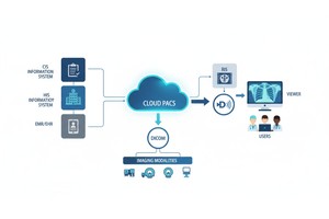
Cloud platforms for imaging help to build this foundation through:
• Web-based Access To Dicom Studies
• Quick Exchange Between Locations And Specialists
• Managed Storage With Process Continuity
• Powerful Infrastructure For Sophisticated Rendering Tasks
• Simpler Team Collaboration For Remote Healthcare
Platforms like PostDICOM already support the new web-based imaging workflows that are in line with the trend towards connected and visualization-driven healthcare.
The move to VR does not have to happen all at once to start preparing for it. They can take many of the necessary steps today by upgrading their imaging systems.
These include secure web-based viewers, remote viewing, seamless collaboration across sites, centralised image management, and systems that are cloud-ready. These changes improve today's workflows and lay a foundation for advanced visualization software of the future.
| Feature | Traditional 2D Review | Advanced Visualization |
| Image Format | Slice-by-slice | Interactive 3D environment |
| Spatial Understanding | Manual interpretation | Immediate visual context |
| Surgical Planning | Limited | Stronger support |
| Training Use | Standard teaching | Immersive simulation |
| Collaboration | Screen sharing | Shared interactive review |
| Workflow Reach | Local workstation focused | Web-enabled and scalable |
New visualization tools can provide valuable operational and clinical benefits.
Manipulation can save time spent mentally integrating anatomy from multiple slices.
3D models are often more readily understood by clinicians.
3D review environments may enhance retention and preparation for procedures.
Visual anatomy may help patients conceptualize and understand medical diagnoses and treatment options.
The cloud allows consultations to take place across multiple workstations and hospital sites.
While the future is promising, successful adoption requires practical planning.
Healthcare organizations should consider:
• Hardware Purchase And Maintenance
• Staff Training Requirements
• Pacs, Ris, Ehr, And Workflow Integration
• Data Privacy And Regulatory Compliance
• Clinical Validation For Specific Use Cases
• Network Performance And Infrastructure Readiness
The best implementations are those where technology helps support and improve workflows, rather than just being new.
DICOM has always been more than a file format. It's the key that enables imaging innovation to be shared across manufacturers, hospitals, and care settings.
As new methods of visualization emerge, DICOM data sets will increasingly support:
• Ai-assisted 3d Interpretation
• Remote Collaborative Diagnostics
• Immersive Surgical Planning
• Cross-platform Mobile Access
• Next-generation Education Environments
And those who invest in new imaging infrastructure can now keep pace as these technologies evolve.
Yes. Several centers are using VR for surgical planning, teaching, anatomy, and some complex imaging procedures.
Augmented reality is the display of digital imaging data superimposed on the real world for planning, guidance, education, and communication.
Yes. It is possible to render CT and MRI scans into interactive 3D models using appropriate software.
Cloud-based systems increase accessibility, sharing, scalability, and collaboration, which are helpful features for advanced visualization.
No. The technology is meant to assist radiologists and speed up the process, not take the place of expertise.
The most common applications are in surgery, cardiology, oncology, orthopedics, neurology, and education-focused institutes.
New visualization tools are giving clinicians a different way to view medical images. As virtual reality, augmented reality, and advanced 3D rendering technologies evolve, doctors and nurses will have quicker, more intuitive, and more collaborative methods for reading complex studies.
This shift won't be instantaneous, but it is clear: the future of imaging is no longer just on a flat screen. Institutions that upgrade their imaging systems now will be able to embrace new diagnostic, planning, and collaborative imaging technologies in the future.
|
Cloud PACS and Online DICOM ViewerUpload DICOM images and clinical documents to PostDICOM servers. Store, view, collaborate, and share your medical imaging files. |