Medical imaging is more than just taking a scan. A CT, MRI or ultrasound image must also be stored, opened, shared, reviewed and linked to the patient and study data. This is achieved in many imaging devices and healthcare systems through the use of the standard, DICOM.
Knowing about DICOM can help hospitals, imaging centers, radiologists, physicians, students, and patients gain a deeper insight into how medical images are exchanged in today's health care environment.
DICOM is the acronym for Digital Imaging and Communications in Medicine. The standard for storing, transmitting, viewing and handling medical imaging data.
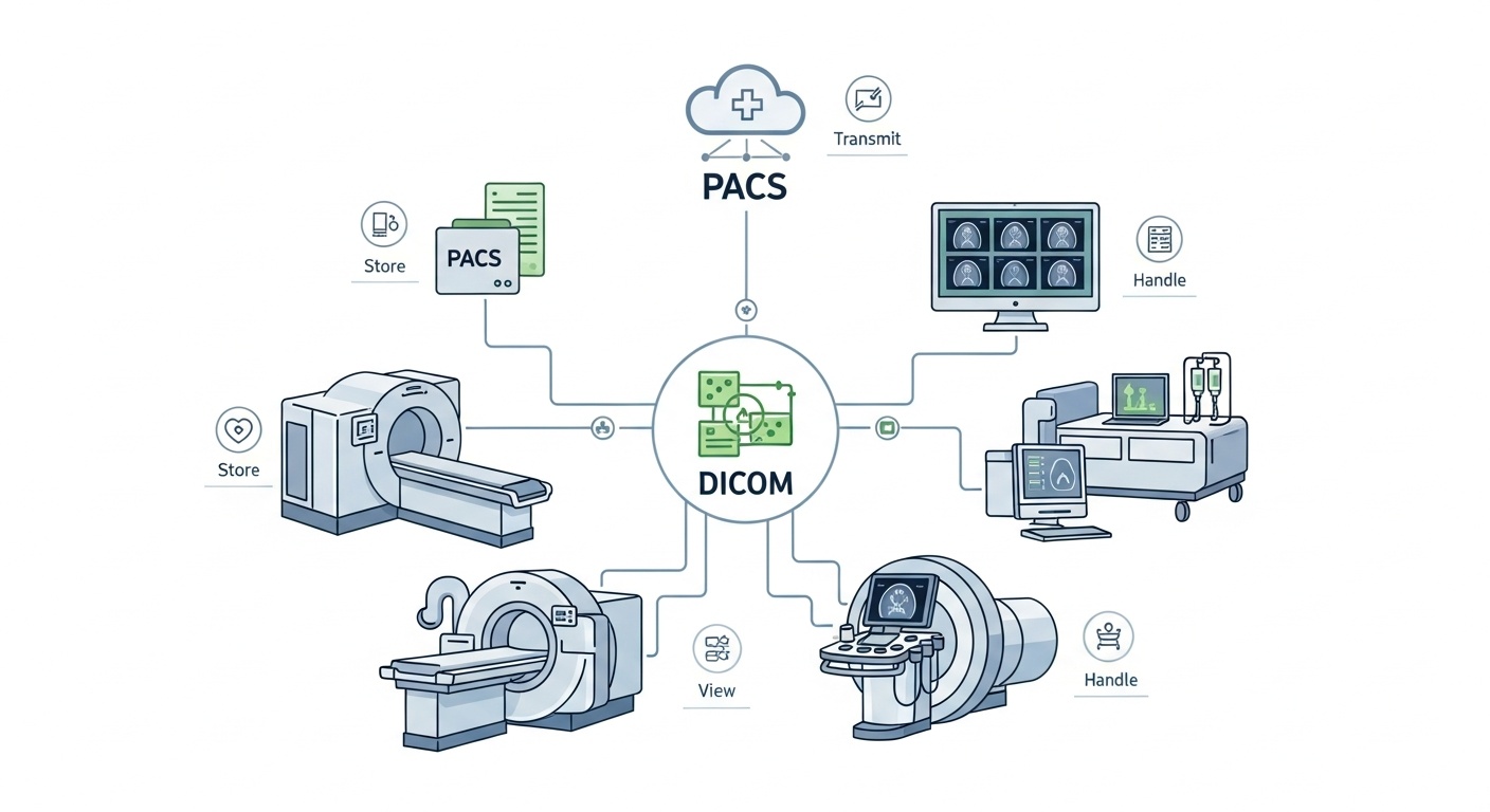
Put simply, DICOM enables medical imaging devices, PACS systems, DICOM viewers and healthcare networks to communicate with one another. Typically, a DICOM file includes the medical image along with pertinent patient, study, modality and image acquisition information.
DICOM is not a diagnostic tool. Rather, it facilitates the imaging process by ensuring that medical images and their related data can be accessed, displayed and shared in a well organized manner.
DICOM is not just a file format, but also a communication standard. This implies it specifies the way in which medical images are stored and how imaging systems communicate with one another in relation to medical images.
For instance, an MRI scanner generates a study, which is typically stored as a DICOM file. These files can be transmitted to a PACS, viewed using a DICOM viewer, stored for future reference, or accessed by another authorized healthcare provider.
The DICOM format is not like JPEG or PNG; it can contain both the medical metadata related to an image and the image itself. These meta data can range from patient ID, study date, modality type, image orientation, slice thickness, body part examined, and more technical data. These details are typically stored in the DICOM tags that aid imaging systems in reading and displaying the image properly.
There are numerous medical imaging systems. One facility might order a scan for a patient, the radiologist might then examine the scan at a different facility and then it might be sent to a referring physician or specialist. In the same way DICOM assists these systems work together with a common structure.
DICOM is advantageous in that it provides the following:
• Improved Device-software Compatibility
• Easier Storage And Retrieval Of Imaging Studies
• More Uniform Presentation Of Images Across Compatible Platforms.
• Collaboration Among Different Departments Or Facilities.
• Use Of Key Image And Study Data Metadata Preservation.
• More Efficient Imaging Workflows For Imaging Teams.
In the radiology and cardiology areas, DICOM is used in medical education, in emergency situations, to plan surgery, to access medical images, in oncology, and more.
DICOM, PACS and DICOM viewers are related and similar but not synonymous.
| Term | What it means | Main role in imaging |
| DICOM | Medical imaging file and communication standard | Stores and transfers images with metadata |
| PACS | Picture Archiving and Communication System | Stores, organizes, retrieves, and manages imaging studies |
| DICOM Viewer | Software used to open and review DICOM images | Allows users to view, measure, and analyze images |
| RIS/EHR | Healthcare information systems | Manage orders, reports, patient records, and clinical workflows |
The difference is easiest to explain as follows: DICOM is similar to the standard, PACS is the storage and management system while a DICOM viewer is the tool for accessing images within the PACS.
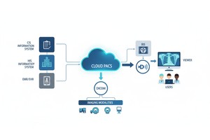
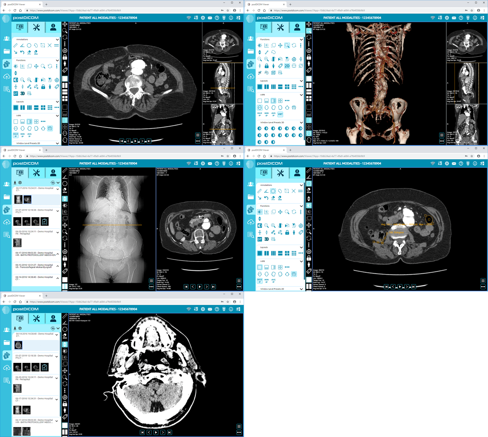
For instance, a modern cloud PACS can store and manage DICOM studies online, and a Diagnostic DICOM Viewer can allow users to access the studies from a compatible workstation or browser.
 - Created by PostDICOM.jpg)
Many medical imaging devices and systems share information in DICOM format. Common modalities that are compatible with DICOM are computed tomography (CT), magnetic resonance imaging (MRI), ultrasound, digital X-ray, mammography, PET, SPECT, angiography, radiation therapy planning systems, and dental imaging systems.
It is also employed by PACS platforms, radiology workstations, hospitals, medical image sharing systems and teaching/research environments. The primary advantage is it allows imaging data to flow between stages of the imaging workflow without losing the image or associated information.
Typically a DICOM file has two sections, the image data and the metadata.
The image data is the actual medical image. The metadata contains information about that image including study information, patient information, modality type, scan parameters, image position, and/or technical settings.
This is one advantage of DICOM over a standard image file for medical imaging. A JPEG can contain an image but is typically not accompanied by detailed clinical and technical context like would be found in medical imaging workflows.
DICOM is used in hospitals and imaging centers to link scanners together with PACS systems, reporting systems and viewers. This facilitates the team to store new research, access old exams and give access to authorized users.
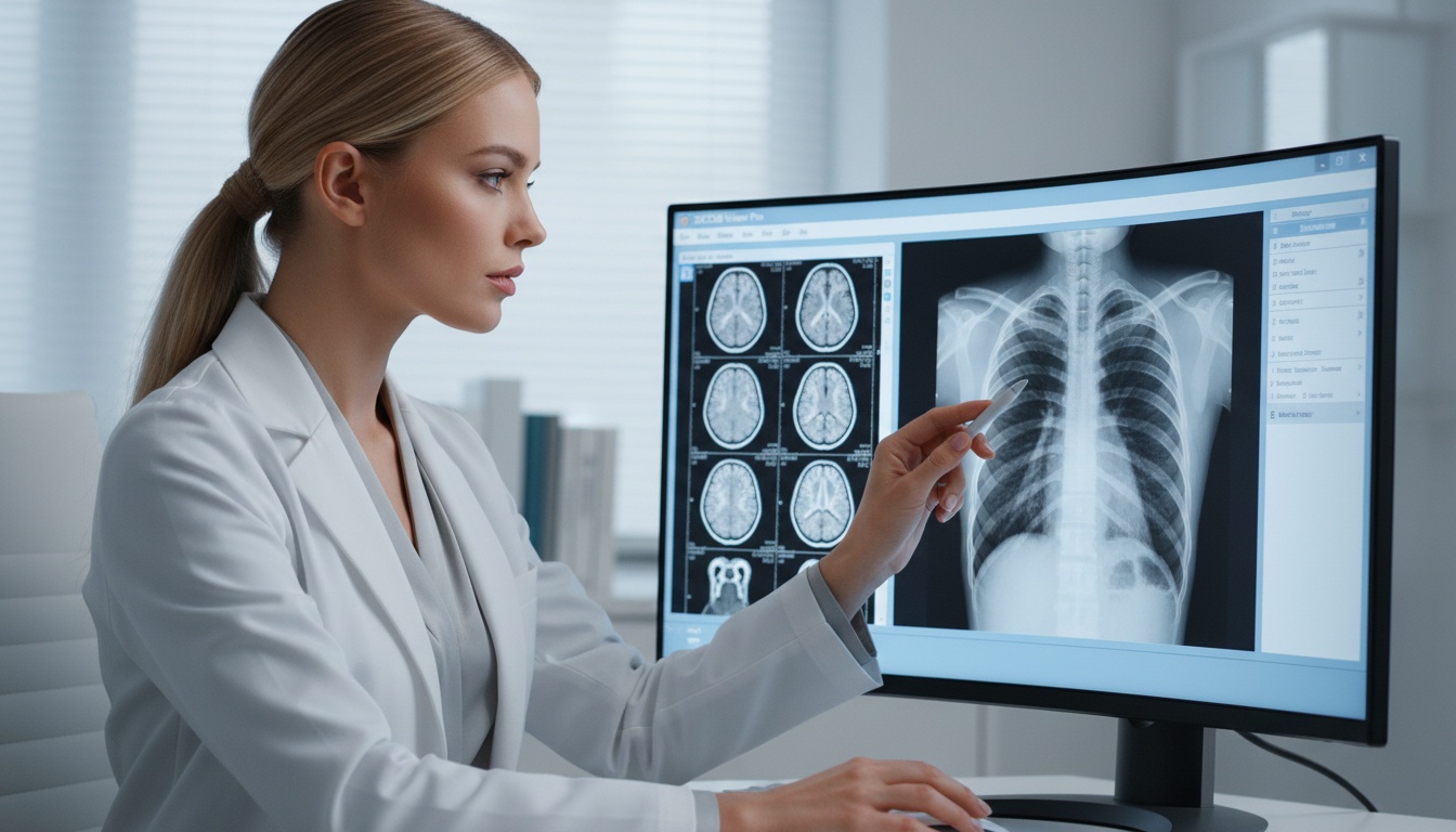
A DICOM viewer is a tool that radiologists use to view imaging studies, compare previous exams, make measurements, and aid in their reporting. DICOM images are viewed in conjunction with other patient information, including reports, by referring physicians.
Imaging studies can be sent to patients via CD, USB drive, patient portal or online-sharing systems. Depending on the viewer or platform, they can open or share their images with another doctor to consult or further treatment.
DICOM can also be applied in the field of teaching and research. Imaging cases can be de-identified for teaching and research purposes, as appropriate.
A typical DICOM workflow starts when a patient undergoes an imaging exam. The scanner generates DICOM images and those images are either pushed to a PACS or cloud imaging system. A radiologist accesses the study in a DICOM viewer, examines the images and writes a report. The images and report can then be sent to the referring physician or another specialist, if necessary.
This process can occur at various sites in today's health care system. A scan may be done at one clinic, read remotely and sent to a specialist at a different clinic. Secure medical image sharing can help decrease the use of physical CDs and make it easier for authorized users to access images.
In DICOM, the data is structured and exchanged in a certain way, but its security rests on how it's implemented. Common requirements include access control, audit logs, privacy protection, encrypted transmission, and secure storage that are typically required for healthcare organizations.
All DICOM workflows should be set up per local laws, regulations, policies, and patient privacy.
The DICOM is Digital Imaging and Communications in Medicine. It's the standard for storing, transmitting and managing medical imaging data.
DICOM is both. It specifies the organization of medical imaging files and the interactions between medical imaging systems.
Yes, but most regular photo viewers cannot properly open DICOM files. You usually need a DICOM viewer or medical imaging platform to view the images and metadata correctly.
Medical images and related data are typically formatted in the standard DICOM. A system for storing, organizing, retrieval and management of DICOM imaging studies is called PACS.
DICOM is chosen because it can store the image and all other important medical metadata together. Although JPEG can present an image, it doesn't typically contain the full context of the study required for medical imaging workflows.
DICOM is an essential standard for medical imaging. It facilitates the exchange of medical images with the appropriate supporting information between imaging devices, PACS, DICOM viewers, and healthcare systems.
DICOM enables healthcare professionals to communicate, exchange images, store images, and access images. Secure systems can simplify the exchange of imaging studies with doctors or specialists for patients.
DICOM is not a diagnosis or treatment tool, per se. It's an industry standard that enables medical images to pass through the healthcare system in a logical and usable manner.
|
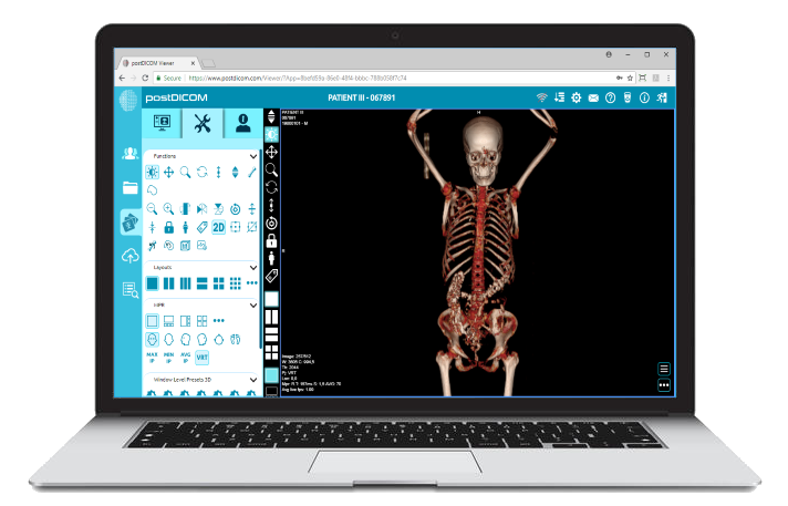
Cloud PACS and Online DICOM ViewerUpload DICOM images and clinical documents to PostDICOM servers. Store, view, collaborate, and share your medical imaging files. |