Medical imaging has advanced significantly from its film-based origins. Today, healthcare professionals rely on digital imaging technologies to diagnose, treat, and monitor patient conditions. At the core of this digital transformation is the DICOM file format: a globally accepted standard for storing and transmitting medical images and associated data.
Whether you're a radiologist, medical IT professional, or simply curious about how your CT or MRI scan is stored and accessed, understanding the anatomy and limitations of a DICOM file is crucial. In this blog, we’ll cover what a DICOM file contains, whether it can hold multiple patient records, and its practical advantages and disadvantages.
We'll also discuss editing, anonymizing, and viewing DICOM files, plus how you can start exploring this format yourself with a free trial from PostDICOM.
DICOM stands for Digital Imaging and Communications in Medicine. It’s both a file format and a network communications protocol developed by the National Electrical Manufacturers Association (NEMA) to ensure seamless interoperability between imaging equipment (such as MRIs, CTs, and X-rays) and software systems (like Picture Archiving and Communication Systems, aka PACS).
Introduced in the 1980s, DICOM was designed to solve a critical problem: the lack of standardization in medical imaging. Before DICOM, different manufacturers used proprietary formats, making it difficult for hospitals to integrate imaging data. DICOM changed the game by introducing a universal standard that embedded both the image and its metadata into a single file.
Today, DICOM is widely used by radiology departments, cardiology units, oncologists, and other healthcare professionals. It allows clinicians to view, transfer, and store medical images consistently and securely.
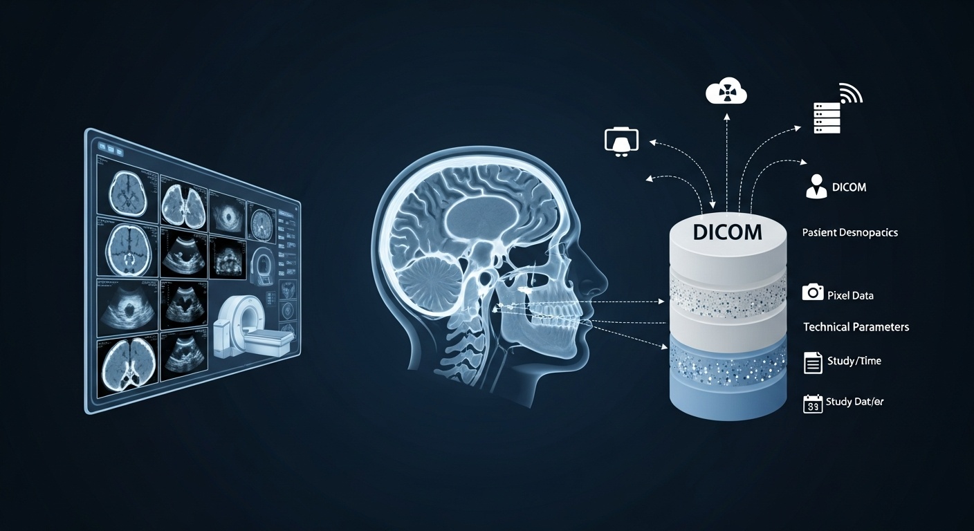
A DICOM file is more than just an image; it contains additional information. It’s a rich container of data that encapsulates everything needed for accurate image interpretation, clinical use, and audit trail maintenance. Each DICOM file consists of two major parts:
This is what makes DICOM uniquely powerful. The header includes:
• Patient Information: Name, ID, date of birth, gender
• Study Details: Modality (CT, MRI, Ultrasound, etc.), study date, referring physician, exam description
• Series Information: Number of images in the series, orientation, body part examined
• Device Information: Scanner model, institution name, software version
These metadata tags are standardized across the DICOM dictionary, which contains over 4,000 unique attributes.
This is the visual part, the actual medical image. Depending on the scan, a DICOM file may contain:
• A Single 2d Image (like A Chest X-ray)
• A Sequence Of Images (like Slices In A Ct Scan)
• A 3d Dataset Reconstructed From 2d Images
Together, the metadata and image data allow doctors to view, analyze, compare, and store patient scans with confidence and traceability.
In DICOM, the Patient ID serves as a key identifier, uniquely linking imaging data to a specific patient within a particular institution or system. According to the DICOM standard, the maximum length of the Patient ID is 64 characters.
That said, many healthcare systems use shorter IDs for simplicity and integration with other systems like EHRs. The ID is case-sensitive, and although alphanumeric values are allowed, special characters should be used cautiously to avoid compatibility issues.
This ID is critical; it connects each image to the correct patient and helps prevent catastrophic mix-ups in clinical workflows.
The short and definitive answer is: No.
By design and according to DICOM standards, a DICOM file is tied to a single patient only. Each file includes metadata fields such as PatientName, PatientID, and PatientBirthDate, which are intended to identify an individual uniquely. Embedding data from multiple patients into a single DICOM file would not only break compliance with the standard, but it would also pose serious ethical, legal, and safety risks.
Why is this rule so strict?
• Privacy: Embedding multiple patients’ data could cause HIPAA (or similar international regulations) violations.
• Clinical Accuracy: Mixing up patient records could lead to life-threatening treatment errors.
• Interoperability: Most PACS systems and DICOM viewers expect one patient per file; violating this requirement can result in file rejection or incorrect labeling.
So, if you ever find yourself dealing with multiple patients’ data, ensure they’re stored in separate DICOM files, even if they’re from the same study or institution.
DICOM is powerful, but like any standard, it has its downsides. Let’s take a closer look:
DICOM is incredibly detailed and flexible, which is great for manufacturers and IT specialists but daunting for new users. The learning curve is steep, especially when working with DICOM tags, transfer syntaxes, and networking protocols.
Even though DICOM is a standard, different vendors sometimes implement features in slightly incompatible ways. This can cause interoperability issues; what works perfectly on one PACS may fail on another.
DICOM files can be massive, especially for CT or MRI studies. This affects storage, transfer time, and system performance. Compression helps, but lossy compression risks degrading image quality.
Unlike JPEGs or PDFs, you can’t just open a DICOM file in a web browser or image viewer. Specialized DICOM viewers are required to interpret both the image and its associated metadata.
If not properly anonymized, DICOM files can expose sensitive patient data. This makes secure handling and sharing essential, especially for research or teaching purposes.
Yes, DICOM files can be edited, but with caution.
There are specialized tools and libraries (such as DCMTK, GDCM, or commercial platforms like PostDICOM) that allow users to:
• Modify Metadata Tags (e.g., Patient Name, Study Description)
• Change Pixel Values (though This Is Rare And Tightly Regulated)
• Add Or Delete Attributes
• Anonymize Or Pseudonymize Patient Data For Research Purposes
However, editing should always preserve the integrity of the original study. In clinical settings, unauthorized edits could result in audit failures or clinical liability. Many institutions maintain an unaltered original copy and use a separate version for teaching or AI training.
 - Created by PostDICOM.jpg)
Anonymization is the process of removing identifiable patient data from DICOM files, which is crucial when files are used for:
• Research
• Education
• Ai Model Training
• Case Sharing Across Institutions
Typical fields removed or modified include:
• Patient Name
• Patient Id
• Date Of Birth
• Referring Physician
• Institution Name
Many DICOM viewers and PACS systems include built-in tools for anonymization. For example, PostDICOM offers streamlined anonymization features that enable you to safely share or export studies without exposing sensitive data.
There’s also the DICOM Supplement 142 guideline, which formalizes anonymization processes, making it particularly beneficial for research institutions seeking compliance.
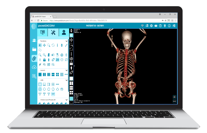
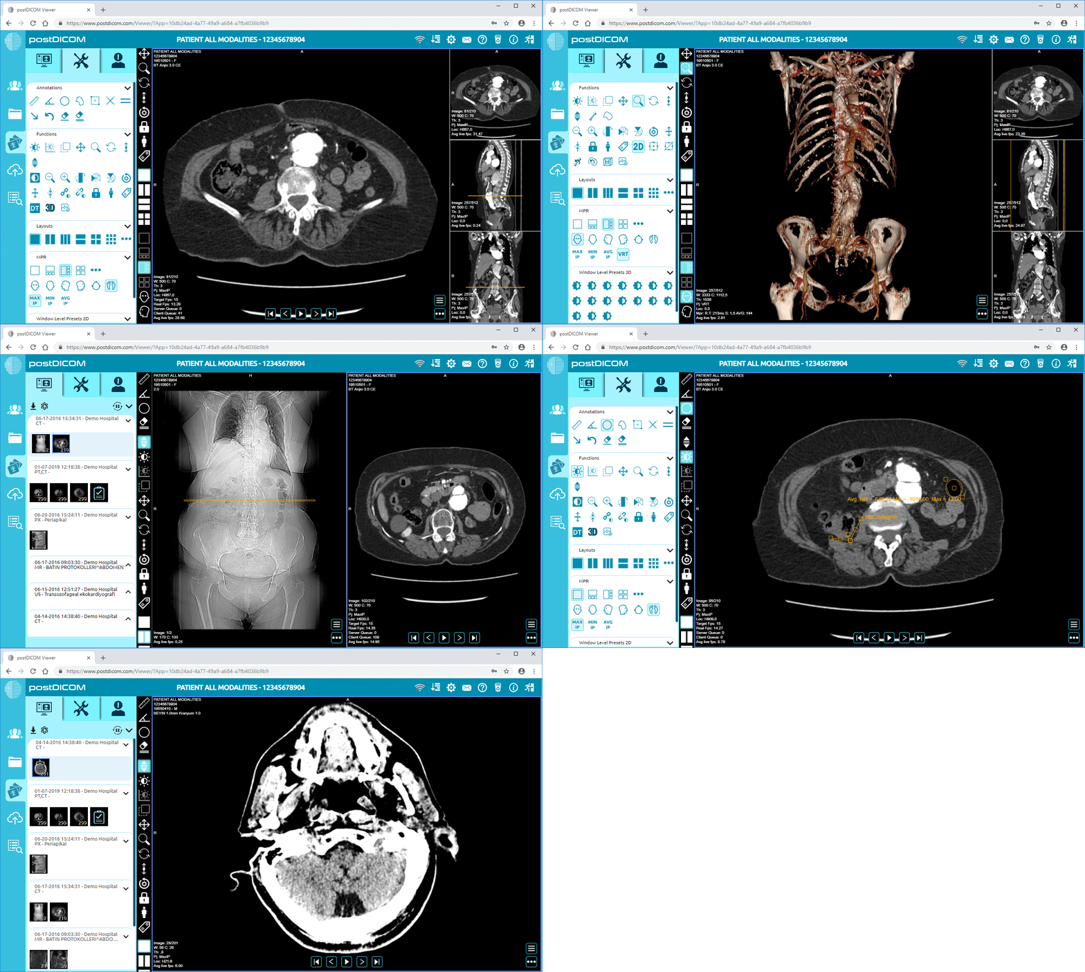
Opening a DICOM file requires more than just an image viewer. It needs software that can interpret both pixel data and metadata.
Here are popular options:
• Postdicom– Acloud-based Dicom Viewerthat’s Intuitive, Powerful, And Great For Professionals And Students Alike.cloud-based DICOM viewer
• Radiant Dicom Viewer– Lightweight Desktop App For Windows
• Horos (mac)– Open-source Viewer Popular In Academic Settings
• Microdicom– Windows-based Viewer With Basic Editing Features
• Weasis– Java-based Viewer, Open-source, Highly Configurable
Of these, PostDICOM stands out thanks to its web-based interface, cloud storage integration, and support for multi-device access. Whether you're a student, radiologist, or healthcare administrator, PostDICOM offers a straightforward way to view, share, and manage DICOM files (no installation required).
DICOM files are at the heart of modern medical imaging. They’re comprehensive digital records that ensure accuracy, traceability, and interoperability across healthcare systems. While a DICOM file can hold multiple images from a study, it is strictly limited to a single patient’s data.
Though powerful, DICOM isn’t without its challenges: it’s complex, sometimes bulky, and requires specialized tools for access, editing, or anonymization. But with the right software, like PostDICOM, working with DICOM files becomes not only manageable but also efficient and user-friendly.
👉 Try PostDICOM for free today and experience a seamless, cloud-based solution for storing, viewing, and sharing medical images. No downloads. No setup. Just clinical-grade imaging at your fingertips.


|
Cloud PACS and Online DICOM ViewerUpload DICOM images and clinical documents to PostDICOM servers. Store, view, collaborate, and share your medical imaging files. |