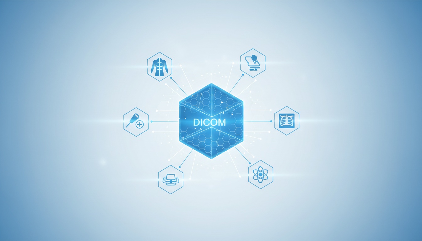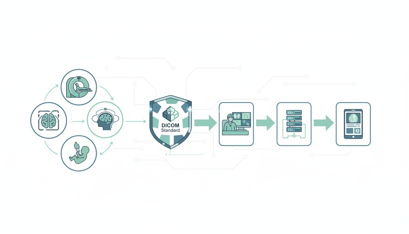
Digital medical imaging has become an integral part of radiology. Today’s radiologist needs to be familiar with the technical aspects of medical imaging. At the forefront of medical imaging technology is the DICOM standard. DICOM stands for Digital Imaging and Communications in Medicine. This is a set of standards framed by the American College of Radiology in association with the National Electrical Manufacturers Association. DICOM ensures that medical images meet quality standards, so that the accuracy of diagnosis can be preserved. All imaging modalities, including CT, MRI and ultrasound must conform to the DICOM standards. Images that are in the DICOM format need to be accessed and used through special DICOM applications. While grasping the basic concept of DICOM is simple enough, the process of actually using DICOM-related hardware and software needs greater technical insight. This article explores the various stages of the DICOM medical image workflow.
DICOM medical images go through different processes—acquisition, storage, editing, secondary processing, sharing and printing, and retrieval. All aspects of this workflow are managed by a central server, referred to as PACS.
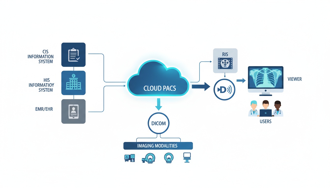
At the heart of the entire DICOM workflow process is PACS. PACS stands for Picture Archiving and Communications System. PACS basically acts as a host that stores DICOM-related imaging data and integrates functions related to these images. You need to think of PACS as the central coordinator where multiple processes meet and integrate. An ideal DICOM PACS software should be able to integrate the source equipment, workstations, sharing networks, retrieving and printing equipment.
In earlier days, medical images acquired by both manual and digital means were printed out as films, or else stored on external devices such as CDs and DVDs. The development of PACS has resulted in a faster and more reliable way of handling medical images. The cost of radiology has decreased because of the reduced need for printed films and hard storage space. Though digital storage was once expensive, the cost has come down considerably over the years. With PACS, the radiologist can manage time better, as images can literally be accessed from anywhere, at any given point of time. The time taken to trace previous images is also reduced. Digital storage of images reduces the chances of inadvertent errors such as mislabelled files and damage to films. The turnaround time to reporting is also lowered because workflow on the whole is enhanced. All this contributes to better patient outcomes.
PACS comprises a number of components essential for the DICOM workflow. These are described below:
The source of all medical images is the hardware equipment that generates them. The detector within the imaging equipment recognizes the image and transmits it in a digitized format to the computer on which it can either be viewed at once or stored for viewing at a later date. All images are acquired and stored in the DICOM format.
|
Cloud PACS and Online DICOM ViewerUpload DICOM images and clinical documents to PostDICOM servers. Store, view, collaborate, and share your medical imaging files. |
Once the images have been acquired from the source, they need to be stored safely. The DICOM server software acts as a filing system to store all images in an organized manner. Images from the DICOM server can either be sent to the digital workstation for interpretation or can be printed. Some advanced DICOM server software applications also allow direct uploading and online sharing of images.
As the name suggests, the DICOM workstation is the area that radiologists use to view and interpret images. The DICOM workstation software is of two types. The first type, called proprietary software, comes along with the source equipment and is usually created by the same manufacturer. This DICOM workstation software must necessarily be used at the same location as the source equipment. The second kind of DICOM workstation software is third-party software, which can be used at a location remote to the source equipment. This kind of software is especially useful in hospitals which have a high inflow of patients coming in for medical imaging examinations. Images can quickly be acquired for each patient, and can be accessed and interpreted by the radiologist at a later date. A third-party DICOM application must be capable of receiving DICOM images from the PACS server. Therefore, this would require DICOM receiver software to be built into the application. DICOM receiver software will allow the radiologist to access images from either the PACS server or from external drives such as CD/DVD drives.
A DICOM workstation software, in addition to opening and receiving capabilities, may have additional features that can help the radiologist improve diagnostic accuracy and track progression of diseases. Some of these features include
Image enhancement: DICOM workstation software can allow radiologists to enhance the quality of the images by altering parameters such as brightness, color and contrast. This feature is usually available on even the most basic DICOM viewers.
Image altering and reconstruction: Advanced DICOM workstation software allows users to manipulate the images that have been originally acquired, so that new information can be gained from the medical images. For instance, the images of a particular anatomical region that have been taken in three planes (axial, coronal and sagittal) can be combined to form a three-dimensional reconstructed image. This gives the radiologist better anatomical orientation. The reconstructed image can also be sliced again, at different planes and angles as compared to the originally acquired images. This technique is referred to as Multiplanar Reconstruction (MPR).
Localizing the region of interest: Areas that absorb maximum or minimum amount of energy can be identified as Maximum Intensity projections (MIP) and Minimum Intensity Projections (MINIP). Identifying these areas may help the radiologist see abnormalities quickly.
Generating reports: A few viewers even allow radiologists to generate reports based on their findings and export them to word processors.
Exporting files: Medical images may be used for teaching and learning purposes. Sometimes medical images may also be used in book and journal publications. Medical images in the DICOM format are huge and often cannot be used directly. It is necessary to compress the file and convert it into another imaging format. There are several imaging file formats available. The JPEG format is most commonly used, and DICOM files can be compressed to a very small size using this format. However, JPEG compression allows data to be lost and the original DICOM file cannot be recovered. Other file formats, like TIFF and PNG, allow recovery of the original file, but they can compress files to only a limited extent. JPEG is useful when one wants to use images in teaching, case presentations, or to present cases on the internet. The TIFF and PNG formats are better suited for book and journal publications.
Anonymizing files: If the medical images are intended for research or are to be published on an open forum such as a website, it is ethically necessary to anonymize the files so that the medical image cannot be linked to a specific patient. Some DICOM applications allow anonymization by removing patient information from the header of the file.
Although printing of DICOM medical images is slowly being phased out, there are still instances where printed films can be useful. For example, when surgical procedures are being performed in remote locations (where the operating complex does not have access to the PACS server), it may be helpful to have the films on display. In these instances, a DICOM printer software may be used to aid in printing films from stored DICOM images. Apart from the DICOM printing software, a printer that is capable of printing DICOM images may also be required.
1. Web-based PACS
With the advent of cloud-based storage, web-based PACS has become a reality. PACS need not be limited to a particular hospital or clinic. All medical images that are acquired at source can be uploaded on to the internet and stored in cloud. This has led to the development of a new off-shoot of radiology—teleradiology. Teleradiology allows radiologists to view images and provide diagnoses at remote centers.
2. Integration of other information systems with PACS
Apart from PACS, there are several other information systems that are in use in hospitals. The Radiology Information System (RIS) is designed to receive orders for medical imaging, generate bills for the same, and to deliver the final report as interpreted by the radiologist. The Hospital Information System (HIS) usually contains the patient’s complete electronic medical records and billing information. More advanced software systems enable integration of all the above information systems with PACS. This allows a complete record of patient information to be stored and made accessible to the clinician, who can then make better informed decisions regarding the medical care of a patient.
To adequately cover the entire DICOM-PACS workflow, you need software that will help you receive the DICOM images from the source (DICOM receiver software), software that will help you view and edit the images (DICOM viewer), software that will help you store all the image data and retrieve it at will (DICOM server software), and software that will help you share and print your medical images (DICOM exporting software and DICOM printing software). Rather than using a multitude of software applications for these purposes, there are programs that fulfil two or more of the above needs. There are, by and large, two kinds of software applications that all radiologists must obtain:
This software primarily allows users to view DICOM images that have been acquired. It usually comes with retrieving and printing capabilities, and some viewers support exporting of files as well. Advanced DICOM viewers have additional workstation features that permit editing and reconstructing of images. A few of the popular DICOM viewers available today include
Horos: Horos is an open-source DICOM viewer that works with Mac OS. The software not only allows viewing but also advanced diagnosis using MPR, MIP and volume rendering. It also has tools for manipulating images and making measurements. Horos also has a plug-in that allows uploading of images to radiopedia, an online resource with a large number of reference cases and articles. Horos cannot, however, integrate to PACS free of cost. It is good for students and residents. Radiologists can use the commercial version called Osirix MD.
RadiANT: The RadiANT DICOM image viewer is a simple, fast platform that is compatible with Windows. It has multiple features, including MPR, MIP and image fusion. Images can be exported to JPEG, PNG and other image formats. They can also be copy-pasted directly into presentations and word documents. The application is just a viewer and does not offer storage space. However, it is useful for students and residents, for studying as well as research.
It is also called DICOM-PACS software and as the name suggests, it has the capacity to store and retrieve DICOM files. A DICOM-PACS software may have its own viewer or it can be integrated to other DICOM viewers. A few examples of free DICOM PACS viewers are
Dicoogle: Dicoogle is an open-source PACS archive that supports storage and retrieval of DICOM files. It has an indexing/query engine that allows users to search for and retrieve DICOM studies. It can work across multiple platforms, including Windows, Linux and Mac OS.
Orthanc: Orthanc is another open-source, lightweight DICOM application. It works across multiple platforms including Windows, Mac OS and Linux, and can turn any computer using one of these operating systems into a mini PACS server. It can also export DICOM data into the PNG format.
SonicDICOM: This is a DICOM server software that also comes with a web-based DICOM viewer. It can be integrated to other DICOM viewers as well. The free version is a trial version and allows up to thirty studies to be saved, with no more than five web or DICOM connections at a time. It is compatible with Windows OS.
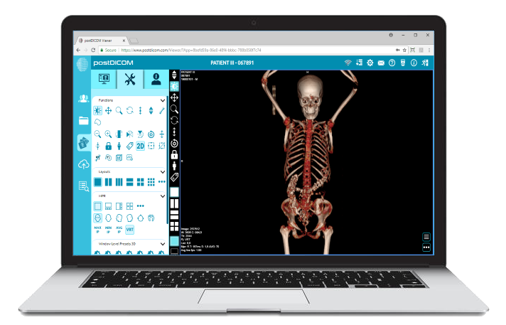
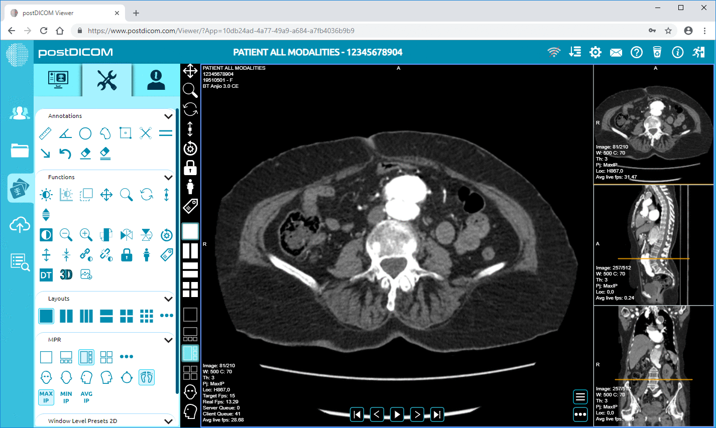
Of note, there are some very handy applications that combine both a DICOM viewer and PACS into a single software. Of these, PostDICOM ranks among the top. It has a powerful, easy-to-use DICOM viewer that offers all advanced imaging features, including MPR, MIP, MINIP and volume rendering. PostDICOM is among the first applications to offer web-based PACS for storing DICOM images. It allows exporting to multiple formats and sharing images is easier through cloud storage. So if you are looking to simplify your DICOM workflow, give PostDICOM a try!
|
|
Cloud PACS and Online DICOM ViewerUpload DICOM images and clinical documents to PostDICOM servers. Store, view, collaborate, and share your medical imaging files. |