Orthopedic imaging is at the center of musculoskeletal diagnosis and treatment. Imaging is a conclusive factor in clinical outcomes, whether it comes to the identification of fractures and joint abnormalities, or the directing of surgical procedures, or the monitoring of recovery.
The last ten years of the century have seen the move towards the adoption of digital radiography as the substitute of analog radiography, which has changed the way in which orthopedic data is recorded. But the actual change is witnessed when the digital X-ray systems are combined with an effective Picture Archiving and Communication System (PACS).
This is not just digitizing images but rather building an interconnected imaging ecosystem, where orthopedic specialists are able to access, analyze and share imaging data in real time. This synergy is being a strategic requirement in the modern healthcare setting, especially when it is incorporating cloud infrastructure, as opposed to a technological enhancement.
In the work of orthopedic practices with growing volumes of patients, trauma cases, and post-surgery visits, this integration has a direct effect on the efficiency of their work and on patient outcomes.
• Pacs With Digital X-ray Can Generate A Very Efficient Orthopedic Imaging Workflow.
• Pacs Can Be Used To Facilitate Quicker Diagnosis, Enhanced Cooperation And Patient Outcomes.
• The Pacs Cloud Is Scalable, Remote And Cost Effective.
• Combination With Health Systems Improves The Overall Clinical Practice.
• The Future Of Ai And Cloud Technology Will Continue To Revolutionize Orthopedic Imaging.
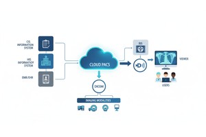
One of the advancements in the orthopedics imaging system is PACS (Picture Archiving and Communication System) which enables the smooth storage, retrieval and exchange of the digital x ray images across departments and locations. PACS could be used together with digital X-ray systems to eliminate the inefficiencies associated with the film, improve the speed of the diagnosis process, remote collaboration, and surgical planning process, and therefore is important in the modern orthopedic practice.
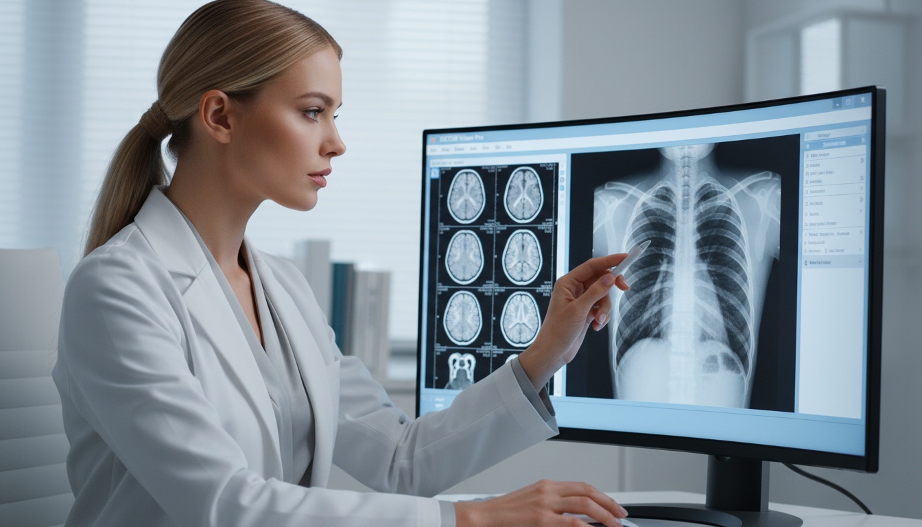
The most popular imaging modality in orthopedics is digital X-ray because it is the fastest, widely available, and cost-effective. It has been used as a first-line diagnostic instrument to assess the bones, joints and alignment problems.
Digital X-ray systems can be used to create images electronically unlike the traditional film based radiography and therefore images can be immediately visualized and manipulated. The orthopedic specialists are able to zoom, contrast, and sharpen certain areas to bring out better diagnostic images.
Digital X-rays are needed in clinical practice to:
• Diagnostic Fractures And Dislocations.
• Assessing Joint Degeneration (e.g., Osteoarthritis)
• Monitoring Post-operative Recovery
• Evaluating The Spinal Positioning And Deformities.
In trauma cases where quick diagnosis is paramount, digital X-ray helps physicians to determine injuries in a few seconds, minimizing time-to-treatment. Nonetheless, although digital X-rays enhance the speed of acquisition and image quality, their maximum benefit is achieved when used with a centralized system such as PACS.
A Picture Archiving and Communication System (PACS) is intended to store, handle, and disseminate medical images within a digital setting. It also requires no physical film archives, and healthcare providers can access imaging information wherever they are, virtually.
In orthopedic processes, PACS forms the foundation of imaging processes by integrating all imaging data into a single system. This will allow clinicians to receive up-to-date and previous research instantly, which is particularly useful during the comparison of pre- and post-operational conditions.
Furthermore, PACS allows collaborating of the radiologists, orthopedic surgeons, physiotherapists, and referring physicians. Such interoperative approach will allow all the stakeholders to have equal access to imaging data, which will assist in eliminating miscommunication and improve coordination of care.
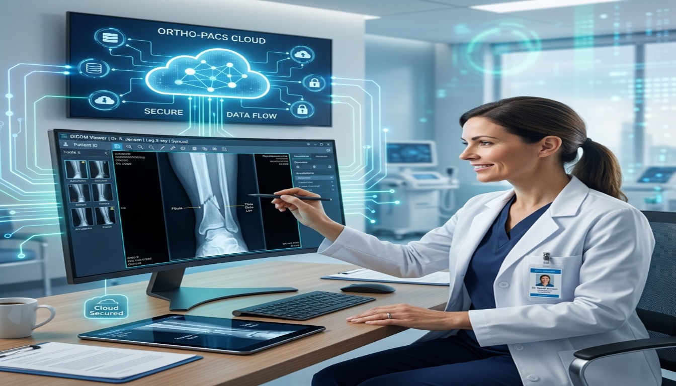
Modern cloud-based PACS systems expand upon these capabilities by enabling access to securely remote access, scalable storage, and real-time collaboration with other facilities, which is essential to the orthopedic practice located on multiple sites.
The digital X-ray systems combined with PACS generated a smooth imaging process, which improves efficiency and accuracy of the diagnostic.
Step-by-step flow:
 - Presented by PostDICOM.jpg)
Such a workflow will remove the time wasted in the process of handling each of them manually, decrease the possibility of duplicating imaging studies, and make sure that clinicians can always access the latest data about the patient.
This workflow is especially useful in orthopedic surgery planning because surgeons will be able to examine imaging data and past research to identify the most effective intervention plan.
 - Presented by PostDICOM.jpg)
To see the actual clinical impact of integration of the PACS, consider a typical case of orthopedic trauma.
The patient presents with an emergency department with a suspected tibial fracture after a sports injury. An X-ray is done digitally and the data is acquired in the DICOM format. The picture is automatically forwarded to PACS system and stored safely and indexed within several seconds.
The radiologist on-call interprets the image with a web based DICOM viewer, initially does an initial interpretation and refers to the case as urgent. At the same time, the orthopedic surgeon (who is not necessarily present in the hospital) also remotely enters into the PACS system and sees the same image in real time.
The surgeon then utilizes PACS to determine the positioning of the fracture, and compare with any prior imaging that is available, and determine whether surgery is necessary or not. This real-time access will allow the care team to make faster and more effective decisions, and treatment delays can be reduced by a significant margin.
Digital X-rays are also performed post-surgery and stored in PACS, which would enable clinicians to monitor how the healing process is progressing over time and renegotiate treatment strategies. This ongoing imaging lifecycle illustrates how through PACS, orthopedic care has turned into a disjointed procedure into a data-driven workflow.
Orthopedic experts can make timely decisions with instant access to imaging data, especially in trauma cases where delays may have a massive effect. The comparison of the current images to the previous studies also increases the accuracy of the diagnosis and minimizes diagnostic errors.
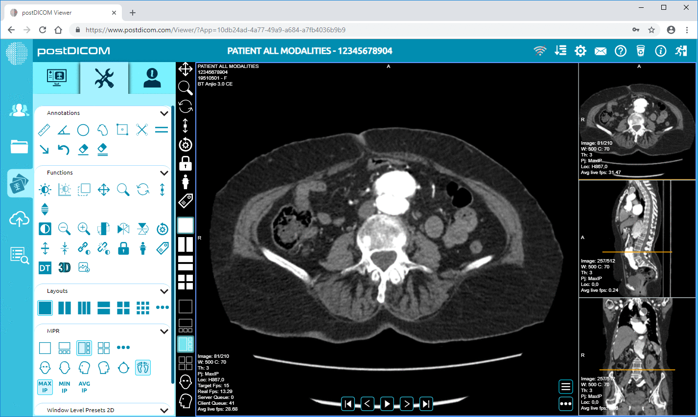
PACS viewers offer sophisticated features like measurements overlaid, annotations and multi-planar imaging. The above features are essential in orthopedic procedures where joint spacing, bone alignment and fracture angles are measured and play a direct role in treatment plans.
The field of orthopedic care commonly requires multidisciplinary cooperation. PACS facilitates easy exchange of images between radiologists, surgeons, physiotherapists and referring physicians, coordinating the management of patients throughout the continuum of care.
There are direct benefits to the patients in faster diagnosis, accuracy and coordinated care resulting in better treatment outcomes, fewer complications and shortened recovery times.
PACS helps to decrease administrative burden by removing both film handling and manual processes. There is an increase in productivity of the imaging departments since they can handle more cases within a shorter period of time.
PACS ensures that long-term expenses related to film, storage, and physical infrastructure decrease even though its introduction might need an initial investment. Also, digital workflows save time and resources because they minimize the necessity of repetitive imaging.
Orthopedic clinics and hospitals can scale their imaging infrastructure easily with cloud PACS, accommodating increasing patient volumes without significant system upgrades.
Cloud PACS allows orthopedic specialists to view images remotely in support of telemedicine and second-opinion consultations. It is particularly useful in the rural or underserved regions where the access to the specialists might be restricted.
With the growth of orthopedic practices and surge in imaging volumes, the conventional on-premise systems tend to lag behind. Cloud PACS is a solution to these constraints because it provides a scalable and dynamic imaging infrastructure.
Cloud PACS allows orthopedic specialists to access imaging data safely regardless of the location, which allows providing consultations faster and managing patients. This specifically comes in handy when practices are located in more than one place or when they associate with outsourced experts.
Technically, cloud PACS systems use distributed storage and content delivery systems so that they can load images quickly, with low latency, even in remote locations. This plays a very important role in the orthopedic processes where huge imaging files should be reviewed immediately.
Also, built-in redundancy and automatic backup guarantees high data availability and disaster recovery mitigating the risk of losing data. The cloud providers are also in charge of updating and patching their systems, which means that the platform is kept up-to-date without the need to utilize internal IT.
| Feature | Traditional Film-Based Imaging | PACS-Enabled Digital Imaging |
| Image Access | Physical retrieval required | Instant digital access |
| Storage | Physical archives | Secure digital/cloud storage |
| Sharing | Manual (films/CDs) | Real-time digital sharing |
| Image Quality | Limited manipulation | Advanced enhancement tools |
| Workflow Speed | Slow and fragmented | Fast and streamlined |
| Remote Access | Not possible | Fully supported |
 - Presented by PostDICOM.jpg)
| Feature | Traditional (On-Premise PACS) | Cloud-Based PACS |
| Infrastructure | Local servers required | No local hardware needed |
| Accessibility | Limited to internal network | Accessible from anywhere |
| Scalability | Limited by hardware | Highly scalable |
| Maintenance | Managed in-house | Managed by provider |
| Cost Model | High upfront cost | Subscription-based |
| Disaster Recovery | Manual backups | Automated redundancy |
To determine the real worth of PACS, one should consider the architecture behind the system in one orthopedic setting.
 - Presented by PostDICOM.jpg)
• Imaging Modalities: Digital X-ray systems generating DICOM images.
• Dicom Gateway: Provides standardization of transmission of images.DICOM Gateway
• Pacs Server / Cloud Storage: Stores and indexes imaging data.
• Viewer Application: Allows clinicians to access and analyze images.
• Integration Layer: Connects PACS with RIS/EHR systems.
In the current cloud systems, the distributed storage, load balancing and secure access protocols to this architecture further help to achieve high availability and performance even when the systems are at peak use. This will make sure that orthopedic teams get access to imaging data that is reliable, whether at another location or not, and whether the system is loaded or not.
Although PACS has great advantages, it must be planned carefully to implement.
Integrating PACS with existing hospital systems (RIS/EHR) may be technically complicated and necessitates appropriate configuration and interoperability standards.
Healthcare data should be based on strict regulations. Cloud PACS providers are to provide encryption, access management, audit history, and regional compliance.
It can be cost-effective in the long term, but small practices moving off old systems may have issues with initial set-up costs.
Personnel should be adequately trained on PACS use. Organizations can also fail to fully enjoy the benefits of the system without proper training.
PACS ought to be adopted by orthopedic practices as they start to witness augmented imaging volumes, workflow unproductivity, or the desire to access and collaborate remotely.
It is especially helpful in case of:
• Multi-location Orthopedic Clinics.
• Acute Care Hospitals With Large Imaging Volumes.
• Telemedicine Consultation Practices.
• Plants That Would Like To Modernize And Expand.
To such organizations, PACS is not merely an upgrade, but a strategic investment that improves efficiency, scalability as well as the quality of patient care.
Artificial intelligence is also being integrated into PACS platforms to help identify fractures, abnormalities, and subtle patterns, which might be missed during manual analysis.
Cloud PACS is fast gaining momentum as the new standard that has provided flexibility, scalability, and accessibility to healthcare providers worldwide.
The use of technologies like 3D reconstruction and high imaging analytics is enhancing surgical planning and accuracy.
The enhanced standards of interoperability are facilitating smooth integration of imaging systems with the larger healthcare ecosystems.
PACS is a digital medical image repository, management, and sharing system that can work faster and enhance collaboration in orthopedics care.
Digital X-ray is a quick and high quality imaging that is necessary in the diagnosis of fracture, joint and musculoskeletal disorders.
PACS removes the manual tasks, provides an opportunity to access images immediately, and promotes effective communication among medical workers.
No, cloud-based PACS enables clinicians to access and review images remotely to support telemedicine and second opinions.
Cloud PACS is highly scalable, can be accessed remotely, and is less expensive to maintain than on-premise systems.
Some of the main trends are AI-based diagnostics, cloud computing infrastructure, advanced visualization and interoperability of the systems.
Cloud PACS systems implement sophisticated encryption, access management, and compliance systems to provide patient data security.
PACS is most suitable in hospitals, multi-location clinics and practices that deal with large volumes of imaging or collaborate remotely.
The use of PACS together with digital X-ray systems is a big step towards orthopedic imaging. It converts disjointed workflows into integrated, data-driven workflows that become more efficient in clinical practice and patient care.
Digital and cloud-based healthcare ecosystems are optimally situated to transform orthopedic practices embracing PACS to provide quicker diagnoses, work in groups, and expand their operations. In this regard, PACS is not a technological device, but a structural element of a contemporary orthopedic imaging system.
|
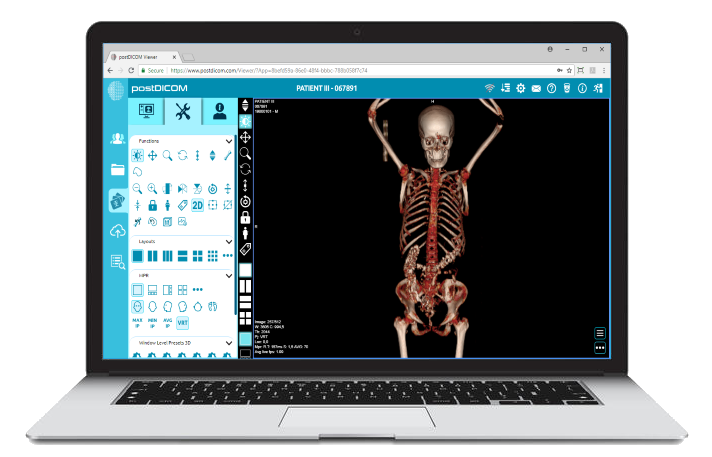
Cloud PACS and Online DICOM ViewerUpload DICOM images and clinical documents to PostDICOM servers. Store, view, collaborate, and share your medical imaging files. |