The moment a mammogram reveals a potential anomaly, the race against time begins in the battle against breast cancer. Integrating mammography with Picture Archiving and Communication Systems (PACS) revolutionizes this critical fight.
This powerful combination is not just an advancement in medical technology; it's a beacon of hope for early and accurate breast cancer detection.
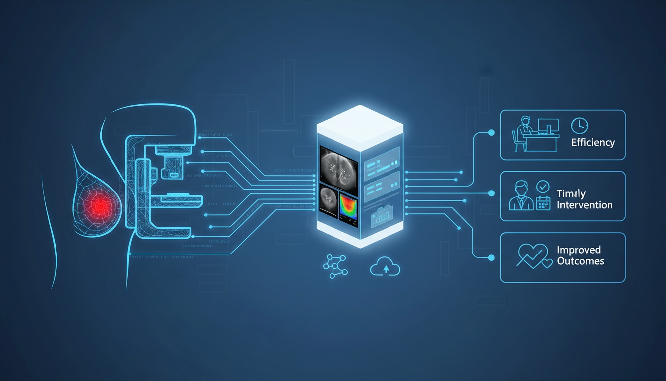
By enhancing image clarity and ensuring immediate access to vital patient data, this integration is reshaping the diagnostic landscape. For healthcare providers, this means more efficient workflows and quicker decision-making.
For patients, it translates to timely interventions and improved outcomes. As we explore this significant development, we'll uncover how the synergy of mammography and PACS sets new benchmarks in breast cancer care, offering insights crucial for those at the forefront of patient care.
The integration of mammography with Picture Archiving and Communication Systems (PACS) is transforming the landscape of breast cancer diagnosis.
This fusion is not just a technical upgrade; it's a significant stride towards more effective, efficient, and secure management of breast cancer imaging.
It's time to see how this integration enhances the quality of mammography imaging, improves storage efficiency, and bolsters data security.
One of the most critical aspects of breast cancer diagnosis is the quality of mammography images. Integrating these images with PACS has significantly improved image clarity and detail.
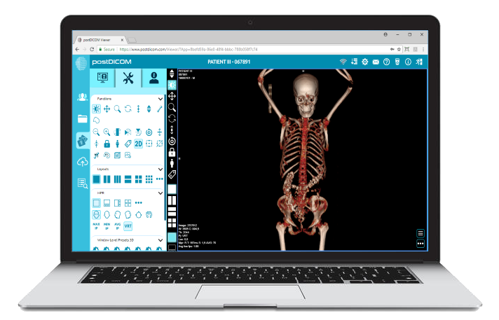
PACS systems often come equipped with advanced viewing tools that allow radiologists to manipulate images for better visualization of breast tissue.
This includes adjusting brightness and contrast, zooming in on areas of interest, and comparing current images with previous ones. Such enhanced image quality is crucial in detecting early signs of breast cancer, which can sometimes be subtle and easy to miss.
Before the era of PACS, mammography images were typically stored on films, which posed challenges in terms of physical storage space and accessibility. With PACS, these images are digitized and stored electronically.
This shift saves physical space and makes retrieval of records quick and easy. Electronic storage means a patient’s mammographic history can be accessed in seconds, facilitating a more comprehensive review of changes over time.
This efficiency is vital in tracking the progression or remission of breast cancer, making PACS an invaluable asset in ongoing patient care.
In the digital age, data security is paramount, especially when it comes to sensitive medical information. Integrating mammography with PACS brings robust security protocols to protect patient data.
These systems are designed to comply with healthcare privacy regulations, such as HIPAA, in the United States, ensuring that patient information is safeguarded against unauthorized access.
Encryption, secure login procedures, and audit trails are standard features of PACS, providing peace of mind for patients and healthcare providers.
The practical impact of integrating mammography with PACS can be seen in healthcare settings across North America. For instance, a breast imaging center in California reported a significant reduction in diagnostic turnaround times after adopting PACS.
This expedited process means quicker treatment planning and potentially better outcomes for patients.
For medical facilities looking to integrate mammography with PACS, the key is choosing the right system that aligns with their needs. Factors to consider include compatibility with existing imaging equipment, ease of use, and vendor support.
Training for radiologists and technicians is also crucial to ensure they can fully leverage the capabilities of the integrated system.
Integrating Picture Archiving and Communication Systems (PACS) with mammography is not just a technological leap; it's a transformation in breast cancer diagnosis that benefits healthcare providers.
From improved diagnostic accuracy to streamlined workflows and enhanced collaboration, let's explore how this integration reshapes breast cancer care in medical facilities.
One of the most significant benefits of integrating PACS with mammography is the substantial improvement in diagnostic accuracy. The enhanced image quality and advanced viewing capabilities of PACS allow radiologists to detect subtle changes in breast tissue more effectively.
For instance, a study conducted at a breast center in New York showed a notable increase in the detection of early-stage breast cancers following the adoption of PACS. This improvement is attributed to the system's ability to provide more explicit images and facilitate a more detailed examination.
The integration of PACS streamlines the entire workflow of mammography imaging. Gone are the days of cumbersome film-based systems.
With digital images stored in PACS, radiologists can access patient scans swiftly, compare current and past images side by side, and make quicker diagnoses.
This efficiency saves time and reduces the patient's anxiety by shortening the wait time for results. A medical center in Toronto reported a 30% reduction in report turnaround time after integrating PACS with their mammography services, significantly enhancing patient satisfaction.
The ability to efficiently retrieve historical patient data is another critical advantage. PACS provides a centralized repository for all imaging data, making accessing a patient's imaging history easy.
This feature is particularly beneficial for patients with a history of breast abnormalities, as it allows for a quick comparison of images over time, aiding in the accurate tracking of changes.
PACS integration extends beyond the confines of a single facility. It facilitates collaboration among healthcare providers, regardless of their location.
Radiologists can share images with specialists for second opinions, discuss complex cases, and make collaborative decisions on patient care. This capability is precious in telemedicine, where specialists can provide their expertise remotely.
For example, a rural clinic in Nebraska utilizes PACS to connect with oncologists in a metropolitan hospital, ensuring their patients receive expert care despite geographical barriers.
Real-world examples underscore the impact of this integration.
A comprehensive cancer center in California integrated PACS with mammography and witnessed a marked improvement in collaborative care, with specialists quickly consulting on cases, leading to more personalized patient treatment plans.
Integrating Mammography with Picture Archiving and Communication Systems (PACS) is not just a technological advancement for healthcare providers; it's a significant stride forward in patient-centered care.
This integration brings numerous benefits impacting patient experience, from faster diagnosis and treatment planning to enhanced data privacy. Let's explore these patient-centric advantages in detail.
One of the most immediate benefits for patients is the expedited diagnosis process. Traditional mammography methods, reliant on physical films, could lead to delays in diagnosis.
With PACS, digital images are instantly available for review, significantly reducing the time from screening to diagnosis. For a patient awaiting mammogram results, this reduced wait time can alleviate anxiety and stress.
Moreover, early diagnosis is crucial in breast cancer treatment, often leading to better outcomes.
For instance, a breast cancer center in Chicago noted a decrease in the time to diagnosis by over 40% after integrating PACS, directly impacting patient prognosis and treatment success.
The integration also streamlines treatment planning. With all imaging data readily accessible, oncologists, surgeons, and other specialists can quickly collaborate to develop a comprehensive treatment plan.
This coordination is vital for complex cases involving multiple treatment modalities. A patient's journey through cancer treatment can be overwhelming, but with streamlined planning, it becomes more manageable and less daunting.
Repeat scans are not just inconvenient; they can also be a source of additional stress and exposure to radiation for patients.
The clarity and detail provided by PACS-integrated mammography reduce the likelihood of inconclusive results, thereby minimizing the need for repeat scans.
A patient at a health clinic in Texas shared how the clarity of her digital mammogram on PACS led to a quick diagnosis, sparing her the anxiety and discomfort of undergoing multiple scans.
Data privacy and security are paramount concerns for patients in the digital age. PACS systems are designed with robust security protocols to protect sensitive health information.
This includes secure data encryption, access controls, and compliance with healthcare privacy laws like HIPAA. Patients can rest assured that their personal health information is safeguarded against unauthorized access, giving them one less thing to worry about during their healthcare journey.
Many PACS platforms offer patient portals where patients can access their medical images and reports. This access empowers patients to be more involved in their healthcare decisions.
Viewing their mammograms and understanding their condition better can lead to more informed discussions with their healthcare providers and greater control over their health.
In conclusion, integrating mammography with PACS is a win-win for patients and healthcare providers. It enhances the efficiency and accuracy of breast cancer diagnosis and significantly improves the patient experience.
From faster diagnoses and coordinated treatment planning to enhanced data security, this integration is a testament to how technological advancements can directly benefit patient care.
As we continue to embrace digital solutions in healthcare, the focus on patient-centric benefits remains paramount, ensuring that every technological step forward is also a step towards better patient care.
 - Created by PostDICOM.jpg)
As we look toward the future of breast cancer diagnosis, integrating mammography with Picture Archiving and Communication Systems (PACS) stands as a beacon of progress.
However, this journey isn't without its challenges. From technical hurdles to financial considerations and the need for specialized training, let's navigate these waters and explore strategies for successful implementation.
The technical aspect is one of the primary hurdles in integrating mammography with PACS. Ensuring compatibility between different systems and software can be daunting.
A key strategy here is to opt for PACS solutions that are known for their interoperability and can easily integrate with various mammography machines and other diagnostic tools.
For instance, a hospital in Seattle overcame these technical challenges by choosing a PACS vendor that offered customizable solutions tailored to their existing infrastructure.
The financial investment in integrating PACS with mammography can be significant.
However, viewing this as a long-term investment in patient care and operational efficiency is essential. Healthcare facilities can explore various funding options to manage financial constraints, including grants, partnerships, or phased implementation strategies.
A clinic in Florida shared how they implemented PACS in stages, allowing them to manage costs without compromising the quality of care.
Effective use of PACS requires specialized training for radiologists, technicians, and other healthcare staff. Continuous education and training ensure that the staff can fully leverage the capabilities of the integrated system.
Investing in comprehensive training programs, online tutorials, and regular workshops can be immensely beneficial. A success story comes from a community health center in Ontario, where a dedicated training program led to a smoother transition and higher adoption rates among staff.
Conduct a Needs Assessment: Before integrating PACS, thoroughly assess your facility’s specific needs and challenges.
Choose the Right Vendor: Select a PACS vendor with scalable solutions and robust customer support.
Involve All Stakeholders: Include radiologists, technicians, IT professionals, and administrative staff in the planning and implementation process.
Prioritize Data Security: Ensure the PACS solution complies with healthcare data security regulations and has robust encryption and access control mechanisms.
Plan for Scalability: Choose a system that can grow with your facility’s needs, accommodate more data, and integrate with new technologies.
The future of breast cancer diagnosis with PACS is not just about overcoming challenges; it's about embracing new opportunities.
With AI and machine learning advancements, we can anticipate PACS systems that offer even more sophisticated diagnostic tools, such as predictive analytics and automated image analysis. These technologies have the potential further to enhance the accuracy and efficiency of breast cancer diagnosis.
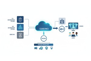
|
Cloud PACS and Online DICOM ViewerUpload DICOM images and clinical documents to PostDICOM servers. Store, view, collaborate, and share your medical imaging files. |