When talking about EEG vs. CT Scan, a common question we see is whether EEG falls under the category of imaging. While diagnosing techniques like CT scans, MRI, and ultrasonography all fall under imaging, we can’t surely say the same thing for EEG. There’s a debate going on as to why EEG may not fall under an imaging modality.
An answer posted by Natalia Murataeva, Doctor of Neuroscience and Psychology, on Researchgate, explained why she thinks EEG is not considered an imaging modality.
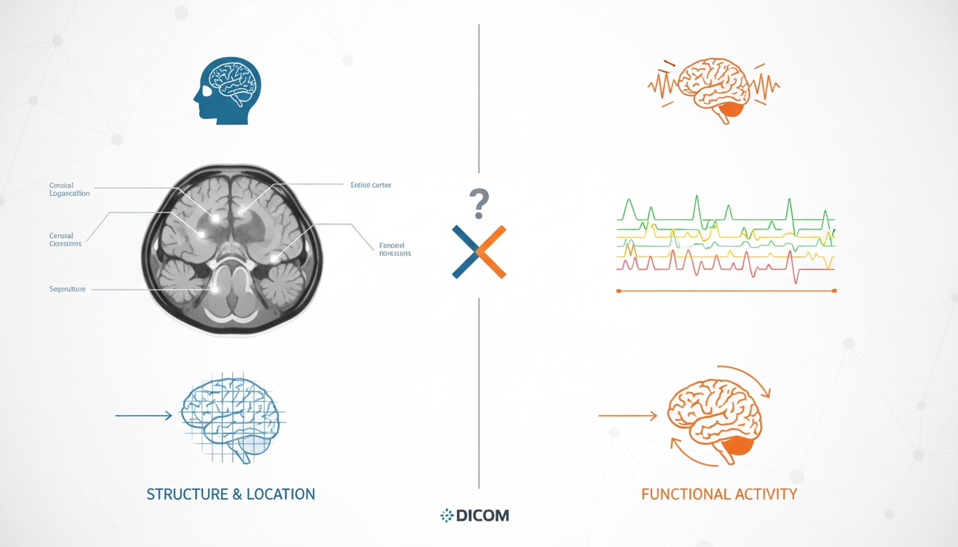
“I think that tomography is an imaging technique and electroencephalography is not because the former provides direct information regarding the structure and its location while the latter provides information only about the functional output of some area. And moreover, the area in question is not well defined either as different people’s information will travel slightly different distances. Hence, no space/shape/form/size info is provided by EEG,” she added.
So, whether EEG falls under image modality or not is still debatable; what we do know for sure is that CT Scan is a part of image modality. Therefore, the best comparison for imaging would be CT Scan vs. MRI, in our humble opinion.
Magnetic Resonance Imaging (MRI) demonstrates structural information of the brain. This information is used to determine the condition of certain areas of the brain. For example, diagnosis of a brain tumor or other abnormal structures.
Electroencephalography, or EEG, is conducted by placing little electrodes on the scalp. The EEG test observes the electrical activity in the brain. The test shows how active the brain is during different activities. For example, diagnosing any anomaly that happened on a specific day.
While the MRI will identify specific “problem areas,” it can’t show any brain activity like EEG. Thus, doctors will most likely recommend other brain imaging methods first. An MRI test is also quite expensive compared to other imaging methods.
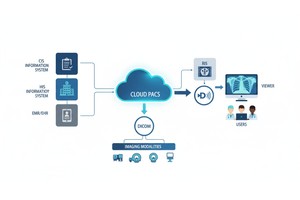
Images like CT scans, MRIs, Ultrasonography, etc., can now be captured, stored, reviewed, and shared with recipients, thanks to DICOM. Digital Imaging and Communications in Medicine, or DICOM, is a standard format for medical images.
The DICOM files contain images and other information about a patient’s diagnosis. These images also have the radiologist’s annotation to understand the report better.
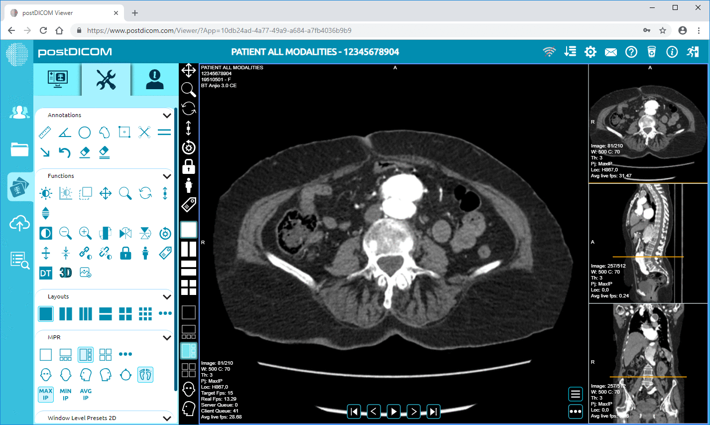
The viewer can manipulate the DICOM data to make simple standard measurements or complex 3D representations of the anatomy. The images can be printed on paper or stored using a cloud storage system called Picture Archiving Communication Systems (PACS). Those who can’t access the system will use CD-ROM with viewing software like PostDICOM to view the files.
Before you proceed to check your CT scan image, keep 3 shades (Gray, Black, and White) in mind as they play a crucial part in the scans. Here is what these 3 shades represent:
Gray: Areas with fluid, soft tissues, and blood will appear in shades of gray.
Black: Areas with fat and air will show up as dark gray or black.
White: Areas with bones will show up as white.
If your CT scan identifies an unexpected color in the images, then that can be a sign of an abnormality. Different types of contrast are used to define the body’s structures better. For example, the scan of your brain after you have had a stroke represents something like this -
The bone of your skull will be likely to give off a bright white shade. The tissues around your brain will look gray or black or sometimes both. If you had a stroke, you would find a small portion of white visible around the black and a gray area. That’s the spot where the stroke occurred. This is because that particular area had been deprived of blood flow, and the fluid leaked out of your injured brain cells. The fluid is white in color, shown in the scan.
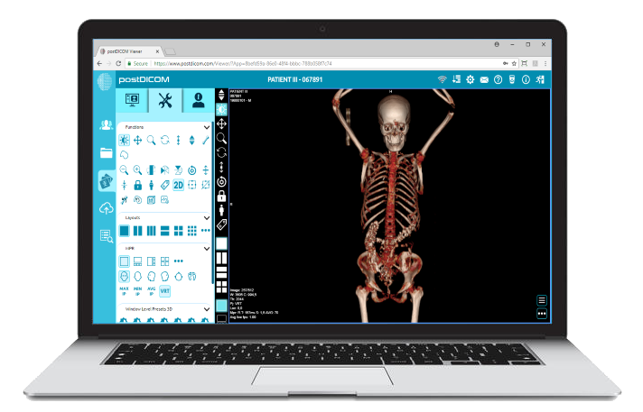
If you’re confused about the right side of the film, observe the words written on the image. Viewing the scan on films is a little trickier than doing it on PACS software like PostDICOM. That’s why doctors, as well as patients, love PostDICOM.
However, if you want to do it manually, here’s how you can do it. First, hold the film in the light. An important thing to note is that the right side of your body will appear on the left side of the film. Similarly, the left side of your body will appear on the right side of the film.
To make it easier for you to read, the uppercase R and L on the films will outline what side of the body is represented in the film. So, for example, the front part of your body will be on the top of the film, and the back part will be on the bottom.
The easiest way to put the films in order is to arrange them according to the numbers printed on the CT films. The CT scan cuts your body into cross-sections. These cross-sections are very thin, just like slices of bread. When you arrange and observe the films in order, you will notice a normal and natural flow. If you notice any sudden breaks in the flow of the films, this may suggest an abnormality.
While viewing so many films at once can be tiresome and adds the chance for mistakes to happen, with PostDICOM’s diagnostic viewer, you can scroll through the images easily. As a result, the images will move in an orderly manner, as if you are watching a slow-moving slideshow.
Now, compare the two sides of your body to help you identify any abnormalities. This one’s especially hard for bilateral organs because they look exactly the same. You can use CT anatomy to compare, but the best way is to compare the scan with the other side.
However, the comparison won’t work for the liver, stomach, and spleen because they aren’t bilateral. On the other hand, you can use the comparison for the two lobes of your brain, your two arms, legs, kidneys, lungs, ovaries, testicles, etc.
Normally, you need to consult with your doctor as soon as you get the reports of your CT scans. The radiologist specializing in interpreting the types of medical images includes a detailed report of what they observed in your scans.
So, we suggest not self-diagnosing and showing your reports to your doctor to understand them better. If you are curious and want to check your report, you can follow the steps we covered. But remember, you can’t master the reading of a CT scan in one day. It takes practice and proper lighting.
DICOM viewers are used for many aspects of medical fields. For example, cardiologists, oncologists, traumatologists, etc., benefit immensely from using DICOM viewers.
The DICOM viewers are constantly changing and improving to meet the demands of professionals and the advancements in medical imaging techniques, for example, CT scans, MRIs, PET-CT, etc.
Specialists constantly need to read the images produced by various medical imaging techniques, and that’s why they need stable viewing and reading software that fits their needs and allows them to work with volumes of information in less time.
Keeping this in mind, PostDICOM has created advanced 3D image-viewing software. Our PACS viewer allows experts to reconstruct images from the given datasets to make a more thorough interpretation that is easy to understand.
Our MPR or Multiplanar reformation is thus very useful for arranging CT scan datasets. In MPR, the data from the complete imaging set is reconstructed to show images in different planes (which weren’t acquired directly during the imaging process).
If we take a CT scan as an example, a regular CT scan dataset only consists of images taken in the axial section. But with MPR, doctors can obtain the coronal, sagittal, and even oblique sections of the body part.
 - Created by PostDICOM.jpg)
Some EEG devices convert their analysis to a PDF file. Then, they can encapsulate that PDF document with DICOM tags to convert it to a DICOM file. Finally, they can send the created DICOM document to PACS systems. PostDICOM Cloud PACS is capable of showing these EEG DICOM analysis results that are in encapsulated PDF format.
Regarding CT scan vs. EEG, it’s unclear if EEG falls entirely under the imaging modality. However, we got to learn how PACS helps in CT scan viewing and reading and why PostDICOM is one of the best software for CT scans.
|
Cloud PACS and Online DICOM ViewerUpload DICOM images and clinical documents to PostDICOM servers. Store, view, collaborate, and share your medical imaging files. |